Paper I — Q1
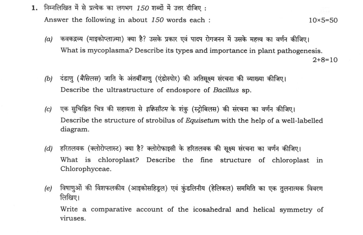
Answer the following in about 150 words each : 10×5=50 (a) What is mycoplasma? Describe its types and importance in plant…
Answer the following in about 150 words each : 10×5=50
What is mycoplasma? Describe its types and importance in plant pathogenesis. 2+8=10
Describe the ultrastructure of endospore of Bacillus sp.
Describe the structure of strobilus of Equisetum with the help of a well-labelled diagram.
What is chloroplast? Describe the fine structure of chloroplast in Chlorophyceae.
Write a comparative account of the icosahedral and helical symmetry of viruses.
हिंदी में प्रश्न पढ़ें
निम्नलिखित में से प्रत्येक का लगभग 150 शब्दों में उत्तर दीजिए : 10×5=50
कवकद्रव्य (माइकोप्लाज्मा) क्या है? उसके प्रकार एवं पादप रोगजनन में उसके महत्व का वर्णन कीजिए। 2+8=10
दंडाणु (बैसिलस) जाति के अंतर्बीजाणु (एंडोस्पोर) की अतिसूक्ष्म संरचना की व्याख्या कीजिए।
एक सुचिहित चित्र की सहायता से इक्विसीटम के शंकु (स्ट्रोबिलस) की संरचना का वर्णन कीजिए।
हरितलवक (क्लोरोप्लास्ट) क्या है? क्लोरोफाइसी के हरितलवक की सूक्ष्म संरचना का वर्णन कीजिए।
विषाणुओं की विषफलकीय (आइकोसाहेड्रल) एवं कुंडलीय (हेलिकल) सममिति का एक तुलनात्मक विवरण लिखिए।
Model answer
Written by UPSC Answer Check against this question's marking rubric, to the 150-word length. UPSC does not publish answers for Mains — this is one way to score well, not an official key.
(a) Mycoplasma: Types and Plant Pathogenesis
Mycoplasmas are wall-less, highly pleomorphic, filterable prokaryotic microorganisms belonging to the class Mollicutes, bounded solely by a trilaminar unit membrane containing sterols. Taxonomically and physiologically, they are categorized into families such as Acholeplasmataceae, which do not require sterols for growth and display non-helical forms, and Spiroplasmataceae, which are sterol-requiring and exhibit helical morphology and rotational motility. Phloem-inhabiting plant forms are classified into Phytoplasmas (non-helical, non-culturable) and Spiroplasmas (helical, culturable).
In plant pathogenesis, these organisms act as obligate parasites restricted to the sieve tube elements of phloem tissue, transmitted biologically by insect vectors such as leafhoppers and psyllids. They induce systemic physiological disorders, floral virescence, phyllody, stunting, and witches'-broom proliferation. In India, phytoplasmas cause economically destructive diseases such as Little Leaf of Brinjal (Solanum melongena) and Sandal Spike Disease (Santalum album), severely disrupting normal shoot architecture and biomass productivity.
**(b) Ultrastructure of Endospore of Bacillus sp.**
The endospore of Bacillus sp. is an extraordinarily resistant, dormant, multi-layered survival structure formed endogenously under nutrient stress. From the exterior to the interior, its ultrastructure reveals:
The exosporium is the outermost, loose, balloon-like membranous layer composed of proteins, polysaccharides, and lipids. Immediately beneath lies the spore coat, a dense, multi-layered proteinaceous shell rich in cysteine and hydrophobic amino acids, providing impermeable protection against lytic enzymes and chemicals. Beneath the coat is the cortex, a thick layer of loosely cross-linked peptidoglycan containing muramic-delta-lactam.
The inner core is bounded by the core wall (germ cell wall), which matures into the vegetative cell wall during germination, and the inner spore membrane, an intact lipid bilayer maintaining extreme impermeability. The innermost region is the core (protoplast), containing the condensed chromosome, ribosomes, and metabolic machinery. The core exhibits extreme dehydration and contains a high concentration of a dipicolinic acid and calcium complex (Ca-DPA), alongside Small Acid-Soluble Spore Proteins (SASPs) that bind and protect DNA from heat, desiccation, and ultraviolet radiation.
`` [ Exosporium ] [ Spore Coat (Outer & Inner) ] [ Cortex (Peptidoglycan) ] [ Core Wall / Germ Cell Wall ] [ Inner Spore Membrane ] [ Core / Protoplast: DNA + SASPs + Ca-DPA ] ``
**(c) Structure of Strobilus of *Equisetum***
The strobilus of Equisetum is a compact, cone-like reproductive structure terminating the main fertile axis or lateral branches. It consists of a stout, central longitudinal axis surrounded by closely packed whorls of specialized reproductive appendages termed sporangiophores, with an anulus forming a protective ring at the base.
``` VERTICAL SECTION OF EQUISETUM STROBILUS ___/___ / Axis \ _______| | |_______ [Peltate [ | | | ] Peltate Disc] [===+===| | |===+===] Disc | | | | | (Sporangia)| | |(Sporangia) [==o==] | | | [==o==] | | | _______| | |_______ [===+===| | |===+===] | | | | | [==o==] | | | [==o==] | | | _________/ [Anulus] || (Fertile Axis)
SPORE STRUCTURE: ( Spore Core )====[ Four Spoon-Shaped Hygroscopic Elaters ] ```
Each sporangiophore consists of a short cylindrical stalk attached at a right angle to the central axis, terminating in an expanded, polygonal, peltate shield-like disc. On the inner, adaxial surface of this peltate disc, 5 to 10 elongated, cylindrical, sac-like sporangia hang downwards parallel to the central stalk. Each sporangium encloses numerous identical, spherical, green haploid spores. Every spore is encircled by four ribbon-like, spoon-shaped, hygroscopic elaters derived from the outer exospore wall and attached at a single point, which uncoil during dry conditions to facilitate spore dispersal.
(d) Fine Structure of Chloroplast in Chlorophyceae
The chloroplast of Chlorophyceae is a specialized, double-membraned photosynthetic organelle showing diverse macroscopic forms, ranging from the cup-shaped plastid of Chlamydomonas to the spiral ribbon of Spirogyra.
Ultrastructurally, it is bounded by a chloroplast envelope consisting of an outer membrane, an intermembrane space, and a selectively permeable inner membrane. Enclosed within is the protein-rich gel matrix termed the stroma, which contains circular plastid DNA (cpDNA), 70S ribosomes, plastoglobules, and enzymes of the Calvin-Benson cycle.
The internal photosynthetic membrane system comprises thylakoids arranged in bands of 2 to 6 appressed membranes forming grana-like stacks, interconnected by unstacked stromal lamellae. Embedded within the stroma are one or more conspicuous pyrenoids, which serve as primary micro-compartments for ribulose-1,5-bisphosphate carboxylase/oxygenase (RuBisCO). Each pyrenoid is enveloped by a distinct sheath of crescentic starch plates, where synthesized starch grains accumulate.
(e) Comparative Account of Icosahedral and Helical Symmetry in Viruses
Viral capsids are constructed through the self-assembly of protein subunits (capsomeres) following two fundamental geometric designs: icosahedral and helical symmetry.
Icosahedral symmetry produces a quasi-spherical, closed isometric shell. The capsid forms a regular polyhedron with exactly 20 equilateral triangular faces, 12 vertices, and 30 edges, operating on a 5-3-2 rotational symmetry (five-fold axes through the vertices, three-fold axes through the face centers, and two-fold axes through the edges). The viral genome is densely packaged within the hollow central interior cavity without directly determining the capsid's outer geometry. Prominent examples include Adenovirus and Cauliflower Mosaic Virus (CaMV).
Helical symmetry produces an elongated, cylindrical, rod-shaped or filamentous structure. The capsomeres are organized in a continuous spiral ribbon around a central rotational axis, forming a hollow central tube. The viral nucleic acid is intimately embedded within an interior protein groove, such that the length of the capsid is strictly governed by the length of the encapsulated genome, and the width is determined by the size and packaging of capsomeres. A classical example is Tobacco Mosaic Virus (TMV).
What "Describe" is asking you to do
Give a full, ordered account of the thing named — its parts, stages or mechanism — in the sequence in which it actually exists or occurs. Most describe questions come from the science optionals, where the marks sit in correct technical detail and, where the stem says so, a labelled diagram.
Structure that answers it
One-line identification of the subject → the parts or stages in their real order, each with its defining detail → labelled diagram where the subject is structural → closing line on function or significance
Where marks are lost
Loose general prose where the examiner is ticking named parts, correct terminology and their sequence; and in the General Studies papers, turning to evaluation before the description is finished.
How this answer will be evaluated
Approach
Framework: Botany Paper 1: Define > Structure/Process > Labelled Diagram > Significance. (a) describe: define > structure or process in order > labelled diagram > significance | (b) describe: define > structure or process in order > labelled diagram > significance | (c) describe: define > structure or process in order > labelled diagram > significance | (d) describe: define > structure or process in order > labelled diagram > significance | (e) describe: define > structure or process in order > labelled diagram > significance Full marks: Precise definitions, detailed structures, clear labelled diagrams, and specific examples.
Key points expected
- Mycoplasma: cell wall-less, phloem-limited, insect vector
- Endospore: core, cortex, coat, DPA
- Equisetum strobilus: sporangiophore, sporangium, elaters, diagram
- Chloroplast: thylakoids, stroma, pyrenoid, Chlorophyceae features
- Virus symmetry: icosahedral (T-number), helical (nucleocapsid), examples
Evaluation rubric
Each sub-part is marked on its own, against the marks and word limit printed on the paper.
- (a) Definition of mycoplasma, classification, and pathogenic mechanism in plants. 10 marks · 150 words
describe— define → structure or process in order → labelled diagram → significance
Must cover
- Define mycoplasma (cell wall-less bacteria)
- List types (MLO, MLO-like, etc.)
- Explain phloem-limited pathogenesis
- Mention vector transmission (insects)
Loses marks
- Confusing with viruses
- Ignoring the 'cell wall-less' feature
Earns more
- Mention specific disease (e.g., Aster yellows)
- Note lack of cell wall
- Mention diagnostic methods (PCR/ELISA)
Extra mark
- Mention specific vector species
- Reference recent biotech control
- (b) Layered ultrastructure of Bacillus endospore from core to coat. · 150 words
describe— define → structure or process in order → labelled diagram → significance
Must cover
- Identify core (DNA, ribosomes)
- Describe cortex (peptidoglycan)
- Describe spore coat layers
- Mention dipicolinic acid (DPA)
Loses marks
- Confusing with vegetative cell
- Missing the cortex layer
Earns more
- Mention germination process
- Note heat resistance mechanism
- Labelled diagram of spore layers
Extra mark
- Mention specific Bacillus species
- Reference industrial application
- (c) Detailed structure of Equisetum strobilus with a labelled diagram. · 150 words
describe— define → structure or process in order → labelled diagram → significance
Must cover
- Describe sporangiophore structure
- Identify sporangium and spores
- Mention elaters (if present)
- Provide a well-labelled diagram
Loses marks
- Unlabelled or missing diagram
- Confusing with other plant cones
Earns more
- Mention heterospory (if applicable)
- Describe the cone axis
- Note the arrangement of sporangiophores
Extra mark
- Mention specific Equisetum species
- Reference fossil significance
- (d) Definition of chloroplast and its fine structure in Chlorophyceae. · 150 words
describe— define → structure or process in order → labelled diagram → significance
Must cover
- Define chloroplast
- Describe thylakoid arrangement (grana)
- Mention stroma and DNA
- Note specific features of Chlorophyceae
Loses marks
- Confusing with other algal groups
- Missing the pyrenoid description
Earns more
- Mention pyrenoid structure
- Describe the envelope membranes
- Labelled diagram of chloroplast
Extra mark
- Mention specific Chlorophyceae species
- Reference photosynthetic efficiency
- (e) Comparative account of icosahedral and helical viral symmetry. · 150 words
describe— define → structure or process in order → labelled diagram → significance
Must cover
- Define icosahedral symmetry
- Define helical symmetry
- Compare capsid structure
- Mention examples for each
Loses marks
- Confusing the two types
- Missing specific examples
Earns more
- Mention T-number for icosahedral
- Describe nucleocapsid arrangement
- Labelled diagrams of both types
Extra mark
- Mention specific virus examples
- Reference vaccine development
Practice this exact question
Write your answer and it is marked point by point against the model answer above — what you covered, what you missed, what you got wrong.
Evaluate my answer →More from Botany 2022 Paper I
- Q1 Answer the following in about 150 words each : 10×5=50 (a) What is mycoplasma? Describe i…
- Q2 (a) What is brown spot disease of rice? Describe its causal organism, symptoms, disease c…
- Q3 (a) What is dikaryotization in fungi? Describe different methods of dikaryotization in ba…
- Q4 (a) What are the characteristic features of the family Orchidaceae? How did the classific…