Paper I — Q6
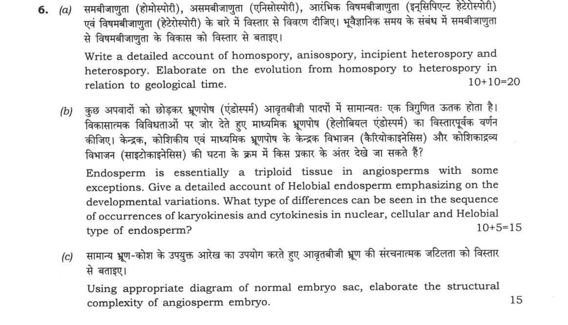
(a) Write a detailed account of homospory, anisospory, incipient heterospory and heterospory. Elaborate on the evolution from…
Write a detailed account of homospory, anisospory, incipient heterospory and heterospory. Elaborate on the evolution from homospory to heterospory in relation to geological time. 10+10=20
Endosperm is essentially a triploid tissue in angiosperms with some exceptions. Give a detailed account of Helobial endosperm emphasizing on the developmental variations. What type of differences can be seen in the sequence of occurrences of karyokinesis and cytokinesis in nuclear, cellular and Helobial type of endosperm? 10+5=15
Using appropriate diagram of normal embryo sac, elaborate the structural complexity of angiosperm embryo. 15 marks
हिंदी में प्रश्न पढ़ें
समबीजाणुता (होमोस्पोरी), असमबीजाणुता (एनिसोस्पोरी), आरंभिक विषमबीजाणुता (इन्सिपिएंट हेटेरोस्पोरी) एवं विषमबीजाणुता (हेटेरोस्पोरी) के बारे में विस्तार से विवरण दीजिए। भूवैज्ञानिक समय के संबंध में समबीजाणुता से विषमबीजाणुता के विकास को विस्तार से बताइए। 10+10=20
कुछ अपवादों को छोड़कर भ्रूणपोष (एंडोस्पर्म) आवृतबीजी पादपों में सामान्यतः एक त्रिगुणित उत्तक होता है। विकासात्मक विविधताओं पर जोर देते हुए माध्यमिक भ्रूणपोष (हेलोबियल एंडोस्पर्म) का विस्तारपूर्वक वर्णन कीजिए। केंद्रक, कोशिकीय एवं माध्यमिक भ्रूणपोष के केंद्रक विभाजन (केरियोकाइनैसिस) और कोशिकाद्रव्य विभाजन (साइटोकाइनैसिस) की घटना के क्रम में किस प्रकार के अंतर देखे जा सकते हैं? 10+5=15
सामान्य भ्रूण-कोश के उपयुक्त आरेख का उपयोग करते हुए आवृतबीजी भ्रूण की संरचनात्मक जटिलता को विस्तार से बताइए। 15
Model answer
Written by UPSC Answer Check against this question's marking rubric, to the expected length. UPSC does not publish answers for Mains — this is one way to score well, not an official key.
The evolution of plant reproduction represents a transition from aquatic dependence to terrestrial independence, marked by increasing complexity in spore production and embryonic development.
Evolution of Spore Types Homospory, the ancestral condition, involves the production of a single type of spore that is morphologically and physiologically identical. In homosporous ferns like Lycopodium and Pteris, these spores germinate into bisexual gametophytes capable of producing both male and female gametes. Anisospory represents an intermediate stage where two distinct spore sizes are produced: microspores and megaspores. While they differ in size, they often lack distinct functional differentiation in early anisospory, though the trend towards functional specialization begins here. Incipient heterospory is a transitional phase seen in certain progymnosperms, where spore size dimorphism is pronounced but the sporangia may not be fully differentiated into distinct microsporophylls and megasporophylls. True heterospory, exemplified by Selaginella and Marsilea, involves the complete separation of microsporangia and megasporangia. Microspores develop into male gametophytes, while megaspores develop into female gametophytes.
Geologically, this transition occurred primarily during the Devonian and Carboniferous periods. The shift from homospory to heterospory was driven by selective pressures favoring reduced gametophyte size and increased parental care. In heterospory, the megasporangium retains the megaspore, providing a protected environment for the developing female gametophyte. This retention of the megaspore within the sporangium is the critical evolutionary step leading to the seed habit, as it allows for the development of a nutritive tissue (endosperm) and a protective coat, ultimately resulting in the origin of gymnosperms and angiosperms.
Helobial Endosperm Development Endosperm is typically a triploid (3n) tissue formed by the fusion of the secondary nucleus (2n) with a male gamete. Helobial endosperm is a common type where the first division of the zygote is followed by a transverse division of the endosperm cell, creating a small chalazal cell and a larger micropylar cell. The chalazal cell typically undergoes further divisions to form a coenocytic or cellular region that often acts as a haustorium, absorbing nutrients from the nucellus. The micropylar cell undergoes free nuclear divisions to form a coenocytic region, which later cellularizes.
Developmental variations exist among taxa. In monocots like Sagittaria, the chalazal region remains coenocytic and haustorial, while the micropylar region becomes cellular. In Eremurus, the pattern is similar but with specific timing differences in cellularization. In dicots like Peperomia, the helobial pattern may show variations in the number of divisions before cellularization.
The sequence of karyokinesis (nuclear division) and cytokinesis (cytoplasmic division) distinguishes endosperm types. In the nuclear type, multiple free nuclear divisions occur before any cytokinesis, resulting in a large coenocyte that cellularizes late. In the cellular type, cytokinesis accompanies every karyokinesis, resulting in immediate cell formation. In the helobial type, the first division is cellular (forming two distinct cells), followed by free nuclear divisions in the micropylar chamber, and finally cellularization.
Structural Complexity of the Angiosperm Embryo Sac and Embryo The normal Polygonum-type embryo sac is a 7-celled, 8-nucleate structure. It consists of the egg apparatus (one egg cell and two synergids) at the micropylar end, a central cell containing two polar nuclei, and three antipodal cells at the chalazal end. The synergids possess a filiform apparatus, a thickened wall structure that guides the pollen tube. Upon double fertilization, one sperm fuses with the egg to form the zygote, and the other fuses with the polar nuclei to form the primary endosperm nucleus.
The structural complexity of the angiosperm embryo itself is derived from the zygote. The zygote undergoes a series of divisions to form the proembryo. In many dicots, the first division is oblique, forming a smaller apical cell and a larger basal cell. The basal cell gives rise to the suspensor, which transports nutrients, and the radicle (embryonic root). The apical cell divides to form the shoot apical meristem and the cotyledons. The embryo is thus a complex, organized structure with distinct regions for future root, shoot, and storage, protected by the developing seed coat and nourished by the endosperm. This intricate organization ensures the survival and establishment of the new plant generation.
What "Elaborate" is asking you to do
Give the full detailed account the question has compressed into a line — every dimension of it, with specifics. Elaborate rewards completeness and detail rather than clarification or argument: the examiner is checking whether you can fill out a topic without being told what its parts are.
Structure that answers it
State the proposition → first dimension in detail → second dimension in detail → the part the statement leaves implicit → the consolidated picture
Where marks are lost
Repeating the statement at greater length instead of adding substance. Elaborate also punishes narrowness: omitting a whole dimension costs more here than anywhere else in this family.
How this answer will be evaluated
Approach
Framework: Botany Paper 1: Define > Structure/Process > Labelled Diagram > Significance. (a) discuss: intro > 3-4 dimensions > example > balanced close | (b) explain: definition/context > points in order > small example > short close | (c) explain: definition/context > points in order > small example > short close Full marks: All parts with labelled diagrams, exact terminology, complete evolutionary/developmental sequences
Key points expected
- Homospory: single spore type, e.g., *Pteris*
- Anisospory: two spore sizes, e.g., *Selaginella*
- Incipient heterospory: transitional stage
- Heterospory: microspores and megaspores
- Geological evolution: Devonian to Carboniferous
- Helobial endosperm: first division transverse, second longitudinal
- Nuclear type: karyokinesis without cytokinesis
- Cellular type: karyokinesis with cytokinesis
- Embryo sac: 7-celled, 8-nucleate structure
- Embryo: cotyledons, plumule, radicle, hypocotyl
Evaluation rubric
Each sub-part is marked on its own, against the marks and word limit printed on the paper.
- (a) Define four spore types and trace their geological evolution. 20 marks
discuss— intro → 3-4 dimensions → example → balanced close
Must cover
- Define homospory, anisospory, incipient heterospory, heterospory
- Evolutionary sequence: homospory to heterospory
- Geological time correlation for each stage
- Labelled diagram of spore types
Loses marks
- Unlabelled diagrams
- Missing geological time context
Earns more
- Examples: *Selaginella*, *Isoetes*, *Psilotum*
- Microspore/megaspore differentiation details
- Adaptive significance of heterospory
Extra mark
- Specific geological periods (e.g., Devonian, Carboniferous)
- (b) Explain Helobial endosperm development and compare karyokinesis/cytokinesis sequences. 15 marks
explain— definition/context → points in order → small example → short close
Must cover
- Helobial endosperm developmental variations
- Karyokinesis vs cytokinesis in nuclear type
- Karyokinesis vs cytokinesis in cellular type
- Karyokinesis vs cytokinesis in Helobial type
Loses marks
- Confusing karyokinesis and cytokinesis
- Missing comparison between three types
Earns more
- Examples: *Oryza sativa*, *Zea mays*
- Labelled diagram of Helobial division
- Triploid nature of endosperm
Extra mark
- Specific species examples for each type
- (c) Use embryo sac diagram to explain angiosperm embryo structural complexity. 15 marks
explain— definition/context → points in order → small example → short close
Must cover
- Appropriate diagram of normal embryo sac
- Structural complexity of angiosperm embryo
- Labelled parts of embryo sac
- Developmental stages of embryo
Loses marks
- Unlabelled diagrams
- Missing structural details
Earns more
- Specific structures: egg cell, synergids, antipodal cells
- Fertilization process details
- Embryo development stages
Extra mark
- Comparison with other plant groups
Practice this exact question
Write your answer and it is marked point by point against the model answer above — what you covered, what you missed, what you got wrong.
Evaluate my answer →More from Botany 2022 Paper I
- Q3 (a) What is dikaryotization in fungi? Describe different methods of dikaryotization in ba…
- Q4 (a) What are the characteristic features of the family Orchidaceae? How did the classific…
- Q5 Answer the following in about 150 words each : 10×5=50 (a) Mention the role of embryo res…
- Q6 (a) Write a detailed account of homospory, anisospory, incipient heterospory and heterosp…
- Q7 (a) Write the uses, botanical name, family and morphology of useful parts of the followin…
- Q8 (a) Describe the protocol involved in rooting and acclimatization of plants produced usin…