Paper I — Q2
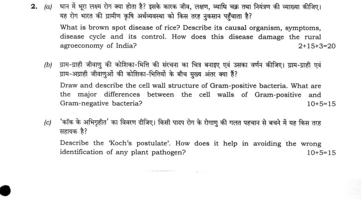
(a) What is brown spot disease of rice? Describe its causal organism, symptoms, disease cycle and its control. How does this…
What is brown spot disease of rice? Describe its causal organism, symptoms, disease cycle and its control. How does this disease damage the rural agroeconomy of India? 2+15+3=20
Draw and describe the cell wall structure of Gram-positive bacteria. What are the major differences between the cell walls of Gram-positive and Gram-negative bacteria? 10+5=15
Describe the 'Koch's postulate'. How does it help in avoiding the wrong identification of any plant pathogen? 10+5=15
हिंदी में प्रश्न पढ़ें
धान में भूरा लक्ष्म रोग क्या होता है? इसके कारक जीव, लक्षण, व्याधि चक्र तथा नियंत्रण की व्याख्या कीजिए। यह रोग भारत की ग्रामीण कृषि अर्थव्यवस्था को किस तरह नुकसान पहुँचाता है? 2+15+3=20
ग्राम-ग्राही जीवाणु की कोशिका-भित्ति की संरचना का चित्र बनाइए एवं उसका वर्णन कीजिए। ग्राम-ग्राही एवं ग्राम-अग्राही जीवाणुओं की कोशिका-भित्तियों के बीच मुख्य अंतर क्या हैं? 10+5=15
'कॉक के अभिगृहीत' का विवरण दीजिए। किसी पादप रोग के रोगाणु की गलत पहचान से बचने में यह किस तरह सहायक है? 10+5=15
Model answer
Written by UPSC Answer Check against this question's marking rubric, to the expected length. UPSC does not publish answers for Mains — this is one way to score well, not an official key.
Brown Spot Disease of Rice
Brown spot is an economically devastating foliar disease of rice caused by the fungus Helminthosporium oryzae (synonym Bipolaris oryzae), whose teleomorph or perfect stage is Cochliobolus miyabeanus.
The symptoms manifest across all aerial plant parts. On leaves, young lesions appear as small, circular, dark-brown specks that enlarge into characteristic oval or spindle-shaped lesions with yellowish halos and greyish-brown centres resembling sesame seeds. On severe infection, lesions coalesce, leading to premature leaf drying. Infection on the panicles and glumes causes dark brown spotting and neck rot, resulting in sterile or discoloured, shrivelled grains.
The disease cycle begins with primary inoculum surviving as dormant mycelium or conidia in infected seeds and crop residues in the soil. Under high relative humidity (above 90%) and warm temperatures (25–30°C), the fungus sporulates, producing multiseptate airborne conidia. These conidia are wind-dispersed, initiating secondary cycles by germinating and directly penetrating the host cuticle or entering via stomata.
Control measures integrate multiple approaches: planting resistant cultivars (including select IR varieties); seed treatment using fungicides like carbendazim or organomercurials to eradicate seed-borne inoculum; cultural management through balanced nitrogen fertilization, optimal plant spacing, and field sanitation; and biological control using Trichoderma harzianum to suppress soil-borne pathogens.
In India, brown spot heavily damages the rural agroeconomy by reducing grain yield and milling quality. It was the primary biological factor behind the 1943 Bengal Famine, where devastating crop losses ranging from 50% to 90% triggered widespread starvation and rural economic collapse.
Cell Wall of Gram-Positive Bacteria
The Gram-positive cell wall is a rigid, thick, single-layered structure (20–80 nm) located outside the plasma membrane. It is primarily composed of multiple layers of peptidoglycan (murein) cross-linked by peptide bridges. Interspersed throughout this network are negatively charged teichoic acids: lipoteichoic acids, which anchor deep into the phospholipid plasma membrane, and wall teichoic acids, which are covalently bound to peptidoglycan.
`` [Outer Surface] | | | | (Lipoteichoic and Wall Teichoic Acids) ===================================================== <-- Thick Peptidoglycan Layer ===================================================== (20–80 nm) ----------------------------------------------------- <-- Periplasmic Space (narrow) ~~~~~~~~~~~~~~~~~~~~~~~~~~~~~~~~~~~~~~~~~~~~~~~~~~~~~ <-- Plasma Membrane (Lipid Bilayer) [Cytoplasm] ``
The major differences between Gram-positive and Gram-negative cell walls include:
- Peptidoglycan: Gram-positive walls contain a thick, multilayered peptidoglycan mesh (20–80 nm), whereas Gram-negative walls have a thin, single-layered peptidoglycan sheet (2–7 nm).
- Outer Membrane: Absent in Gram-positive bacteria, but present in Gram-negative bacteria, where it is composed of an asymmetric bilayer containing lipopolysaccharides (LPS or endotoxins), porin channel proteins, and Braun's lipoproteins.
- Teichoic Acids: Present in Gram-positive walls; completely absent in Gram-negative walls.
- Periplasmic Space: Gram-positive bacteria possess a very narrow, indistinct periplasmic space, whereas Gram-negative bacteria have a prominent, well-defined periplasmic zone containing hydrolytic enzymes and transport proteins.
Koch's Postulates and Plant Pathogen Identification
Koch's postulates provide the classical four-step framework to prove disease causation: First, the specific microorganism must be consistently associated with all diseased individuals and absent from healthy ones. Second, the suspected pathogen must be isolated from the diseased host and grown in axenic (pure) culture. Third, inoculation of the pure-cultured organism into a healthy, susceptible host must reproduce the characteristic symptoms of the disease. Fourth, the identical microorganism must be successfully re-isolated from the experimentally inoculated host.
These postulates prevent the misidentification of plant pathogens by systematically excluding non-pathogenic saprophytes, commensals, and opportunistic secondary invaders that rapidly colonize dying or dead plant tissues. By enforcing pure-culture isolation and artificial re-infection, they distinguish primary causal agents from passive colonizers.
However, standard postulates fail for obligate biotrophs (such as powdery mildews and rusts) that cannot be cultivated on synthetic media, requiring propagation on living host tissue. In modern pathology, molecular Koch's postulates supplement classical methods by using targeted gene disruption to confirm that specific virulence genes are responsible for pathogenicity.
Rigorous application of pathological diagnosis and pathogen isolation remains central to developing targeted crop protection strategies and sustaining national food security.
What "Describe" is asking you to do
Give a full, ordered account of the thing named — its parts, stages or mechanism — in the sequence in which it actually exists or occurs. Most describe questions come from the science optionals, where the marks sit in correct technical detail and, where the stem says so, a labelled diagram.
Structure that answers it
One-line identification of the subject → the parts or stages in their real order, each with its defining detail → labelled diagram where the subject is structural → closing line on function or significance
Where marks are lost
Loose general prose where the examiner is ticking named parts, correct terminology and their sequence; and in the General Studies papers, turning to evaluation before the description is finished.
How this answer will be evaluated
Approach
Framework: Botany Pathology & Microbiology Standard. (a) describe: define > structure or process in order > labelled diagram > significance | (b) describe: define > structure or process in order > labelled diagram > significance | (c) describe: define > structure or process in order > labelled diagram > significance Full marks: Precise definitions, accurate diagrams, specific examples, and clear economic/identification links.
Key points expected
- *Bipolaris oryzae*
- Brown spots, neck rot
- Disease cycle: debris -> spores -> infection
- Control: resistant varieties, fungicides
- Gram-positive: thick peptidoglycan, teichoic acids
- Gram-negative: thin peptidoglycan, outer membrane, LPS
- Koch's postulates: association, isolation, reproduction, re-infection
- Molecular Koch's postulates
Evaluation rubric
Each sub-part is marked on its own, against the marks and word limit printed on the paper.
- (a) Define disease, detail causal agent, symptoms, cycle, control, and economic impact. 20 marks
describe— define → structure or process in order → labelled diagram → significance
Must cover
- Causal organism *Bipolaris oryzae* (or *Cochliobolus miyabeanus*)
- Symptoms: brown spots on leaves, neck rot
- Disease cycle: survival on debris, spore dispersal
- Control: resistant varieties, fungicides, crop rotation
Loses marks
- Confusing with *Magnaporthe oryzae* (Blast)
- Vague economic impact without specific data
- Missing disease cycle steps
Earns more
- Specific fungicide names (e.g., Mancozeb, Propiconazole)
- Quantified yield loss in India
- Impact on rural livelihoods and food security
Extra mark
- Mention of specific Indian rice cultivars
- Recent biotech resistance markers
- (b) Draw Gram-positive wall, describe structure, and list differences from Gram-negative. 15 marks
describe— define → structure or process in order → labelled diagram → significance
Must cover
- Labelled diagram of Gram-positive cell wall
- Thick peptidoglycan layer (20-80 nm)
- Presence of teichoic acids
- Absence of outer membrane
Loses marks
- Unlabelled or missing diagram
- Confusing teichoic acids with LPS
- Incorrect thickness values
Earns more
- Comparison table of Gram-positive vs Gram-negative
- Mention of lipopolysaccharide (LPS) in Gram-negative
- Periplasmic space differences
Extra mark
- Specific peptidoglycan cross-linking details
- Mention of specific Gram-positive species
- (c) State Koch's postulates and explain their role in pathogen identification. 15 marks
describe— define → structure or process in order → labelled diagram → significance
Must cover
- Four postulates: association, isolation, reproduction, re-infection
- Step-by-step process of proving causation
- Application to plant pathogens
- Limitations (e.g., unculturable organisms)
Loses marks
- Missing any of the four postulates
- Vague explanation of 'avoiding wrong identification'
- Confusing with Pasteur's germ theory
Earns more
- Example of a plant pathogen identified via postulates
- Mention of molecular Koch's postulates
- Role in avoiding misdiagnosis
Extra mark
- Specific plant disease example (e.g., *Xanthomonas* in rice)
- Mention of modern molecular techniques
Practice this exact question
Write your answer and it is marked point by point against the model answer above — what you covered, what you missed, what you got wrong.
Evaluate my answer →More from Botany 2022 Paper I
- Q1 Answer the following in about 150 words each : 10×5=50 (a) What is mycoplasma? Describe i…
- Q2 (a) What is brown spot disease of rice? Describe its causal organism, symptoms, disease c…
- Q3 (a) What is dikaryotization in fungi? Describe different methods of dikaryotization in ba…
- Q4 (a) What are the characteristic features of the family Orchidaceae? How did the classific…
- Q5 Answer the following in about 150 words each : 10×5=50 (a) Mention the role of embryo res…