Paper I — Q1
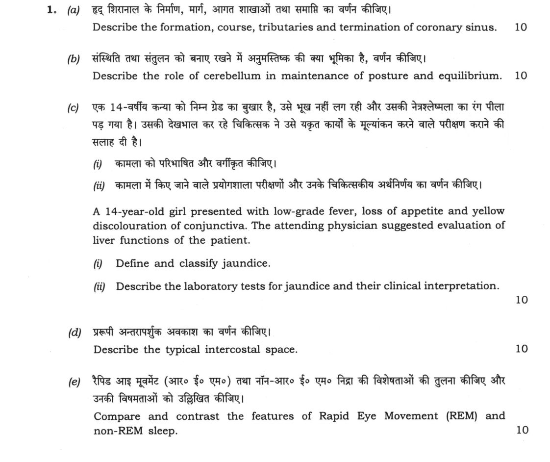
(a) Describe the formation, course, tributaries and termination of coronary sinus. 10 marks (b) Describe the role of cerebellum…
Describe the formation, course, tributaries and termination of coronary sinus. 10 marks
Describe the role of cerebellum in maintenance of posture and equilibrium. 10 marks
A 14-year-old girl presented with low-grade fever, loss of appetite and yellow discolouration of conjunctiva. The attending physician suggested evaluation of liver functions of the patient. Define and classify jaundice.
Describe the laboratory tests for jaundice and their clinical interpretation. 10 marks
Describe the typical intercostal space. 10 marks
Compare and contrast the features of Rapid Eye Movement (REM) and non-REM sleep. 10 marks
हिंदी में प्रश्न पढ़ें
हृद् शिरानाल के निर्माण, मार्ग, आगत शाखाओं तथा समाप्ति का वर्णन कीजिए। 10
संस्थिति तथा संतुलन को बनाए रखने में अनुमस्तिष्क की क्या भूमिका है, वर्णन कीजिए। 10
एक 14-वर्षीय कन्या को निम्न ग्रेड का बुखार है, उसे भूख नहीं लग रही और उसकी नेत्रलेश्मला का रंग पीला पड़ गया है। उसकी देखभाल कर रहे चिकित्सक ने उसे यकृत कार्यों के मूल्यांकन करने वाले परीक्षण कराने की सलाह दी है। कामला को परिभाषित और वर्गीकृत कीजिए।
कामला में किए जाने वाले प्रयोगशाला परीक्षणों और उनके चिकित्सकीय अर्थनिर्णय का वर्णन कीजिए। 10
प्ररूपी अंतरापृष्ठीय अवकाश का वर्णन कीजिए। 10
रैपिड आई मूवमेंट (आर० ई० एम०) तथा नॉन-आर० ई० एम० निद्रा की विशेषताओं की तुलना कीजिए और उनकी विषमताओं को उल्लिखित कीजिए। 10
Model answer
Written by UPSC Answer Check against this question's marking rubric, to the expected length. UPSC does not publish answers for Mains — this is one way to score well, not an official key.
(a) Coronary sinus. The coronary sinus is the principal venous channel draining the myocardium. It is formed in the posterior atrioventricular groove by the union of the great cardiac vein and the oblique vein of the left atrium, also called the vein of Marshall, which is a remnant of the left superior vena cava. From this junction it runs in the posterior atrioventricular groove, between the left atrium and the left ventricle, towards the right atrium. It terminates by opening into the right atrium near the coronary ostium, guarded by the valve of Thebesius. Along its course it receives the small cardiac vein from the right atrium and right ventricle, the middle cardiac vein from the posterior interventricular groove, the left posterior ventricular vein, and several small cardiac veins. The great cardiac vein begins at the cardiac apex in the anterior interventricular groove, ascends, turns into the posterior atrioventricular groove, and becomes the coronary sinus. The anterior cardiac veins drain directly into the right atrium, while most posterior myocardial veins drain through the coronary sinus. This arrangement returns deoxygenated blood from the heart to the right atrium and is clinically important in catheterisation, pacing, and coronary venography. Dilatation of the coronary sinus may be seen in right atrial pressure elevation or anomalous pulmonary venous return. The coronary sinus is separated from the right atrium by a thin wall and may be cannulated via the femoral vein for electrophysiological studies.
(b) Cerebellum in posture and equilibrium. The cerebellum maintains posture and equilibrium by integrating vestibular, proprioceptive, and visual inputs and correcting motor errors. The vestibulocerebellum, comprising the flocculonodular lobe, receives vestibular information through the inferior cerebellar peduncle and projects mainly to the vestibular nuclei and reticular formation via the fastigial nucleus. It adjusts head, eye, and trunk movements to maintain balance, and its output influences vestibulospinal and reticulospinal pathways. The spinocerebellum, especially the vermis and paravermal zones, receives proprioceptive input from the spinal cord through the inferior and superior cerebellar peduncles and modulates tone and posture through the fastigial nucleus to the reticular formation and red nucleus, and through the dentate nucleus to the thalamus and motor cortex. Purkinje cell output is inhibitory and shapes the activity of the deep cerebellar nuclei, allowing fine correction of movement. The cerebellum does not initiate movement but compares intended and actual movement, correcting errors. Lesions of the flocculonodular lobe produce vertigo, nystagmus, and truncal ataxia; vermis lesions cause gait ataxia, wide-based gait, and difficulty with tandem walking, while hemispheric lesions cause limb ataxia. In clinical practice, alcohol, Wernicke encephalopathy, and posterior fossa lesions can impair these circuits. The vestibulocerebellum also contributes to gaze stability and postural reflexes during head movement.
(c) Jaundice. Jaundice is yellow discoloration of the sclera and skin due to serum bilirubin exceeding about 2.5 mg/dL, or 43 µmol/L. It is classified by the site of disturbance: pre-hepatic or haemolytic, hepatic or hepatocellular, and post-hepatic or obstructive. In a 14-year-old girl in India with fever and anorexia, acute viral hepatitis, especially hepatitis A and E, is common, and a hepatocellular cause should be considered until proven otherwise. Serum bilirubin is measured as total, conjugated/direct, and unconjugated/indirect. In pre-hepatic jaundice, unconjugated bilirubin predominates, liver enzymes are normal, alkaline phosphatase and GGT are normal, urine bilirubin is absent, and urobilinogen is increased. In hepatic jaundice, both fractions rise, ALT and AST are markedly raised, alkaline phosphatase and GGT may be moderately raised, viral markers such as HBsAg, anti-HCV, and HAV/HEV IgM when indicated are positive, and prothrombin time/INR may be prolonged in severe disease. In post-hepatic jaundice, conjugated bilirubin predominates, alkaline phosphatase and GGT are disproportionately raised, transaminases are mildly raised, urine bilirubin is positive, and stools may be pale. Conjugated bilirubin is water soluble and appears in urine, causing dark urine. A rise in unconjugated bilirubin suggests increased production or impaired uptake/conjugation, while a rise in conjugated bilirubin suggests impaired excretion or obstruction. Additional tests include complete blood count, peripheral smear, reticulocyte count, serum albumin, and PT/INR to assess synthetic function and haemolysis.
(d) Typical intercostal space. A typical intercostal space is the interval between adjacent ribs, best described in the third to sixth spaces. It is bounded above by the lower border of the rib above, below by the upper border of the rib below, anteriorly by costal cartilage, and posteriorly by the angle of the rib. The space contains the external intercostal, internal intercostal, and innermost intercostal muscles, the pleura, lymphatics, and the intercostal neurovascular bundle. The bundle lies in the costal groove on the inner surface of the lower rib, and from above downward the arrangement is vein, artery, and nerve. The intercostal artery arises from the internal thoracic artery anteriorly and from intercostal arteries posteriorly; the vein drains into the internal thoracic or azygos/hemiazygos system; the intercostal nerve lies below the artery and gives lateral and anterior cutaneous branches. The external intercostal fibres run downward and forward, the internal fibres upward and backward, and the innermost layer is variable. The lower rib protects the bundle, which is why thoracotomy incisions are made over the upper border of the lower rib.
(e) REM and non-REM sleep. Sleep alternates between non-REM and REM stages in cycles of about 90 minutes. Non-REM sleep includes N1, N2, and N3. N1 is light sleep with low-voltage theta activity; N2 shows sleep spindles and K-complexes; N3 is slow-wave sleep with high-voltage, low-frequency delta waves. During non-REM sleep, muscle tone is reduced but present, eye movements are slow or absent, respiration and heart rate are regular, and dreams are less vivid. REM sleep is characterised by low-voltage, mixed-frequency EEG activity resembling wakefulness, sawtooth waves, phasic rapid eye movements, and loss of skeletal muscle tone except the diaphragm. Autonomic activity is variable, with irregular respiration, heart rate, and blood pressure, and vivid dreaming occurs. The first REM period appears after about 90 minutes, and REM duration increases in later cycles. Non-REM sleep is important for physical restoration and memory consolidation, while REM sleep is associated with emotional regulation and procedural learning. Clinically, REM sleep behaviour disorder, narcolepsy, and sleep apnoea illustrate the importance of these stages. Thus, REM and non-REM sleep are distinct but complementary phases that together maintain restorative sleep.
What "Describe" is asking you to do
Give a full, ordered account of the thing named — its parts, stages or mechanism — in the sequence in which it actually exists or occurs. Most describe questions come from the science optionals, where the marks sit in correct technical detail and, where the stem says so, a labelled diagram.
Structure that answers it
One-line identification of the subject → the parts or stages in their real order, each with its defining detail → labelled diagram where the subject is structural → closing line on function or significance
Where marks are lost
Loose general prose where the examiner is ticking named parts, correct terminology and their sequence; and in the General Studies papers, turning to evaluation before the description is finished.
How this answer will be evaluated
Approach
Framework: Medical Science, Paper 1. (a) describe: define > structure or process in order > labelled diagram > significance | (b) describe: define > structure or process in order > labelled diagram > significance | (c(i)) define: precise definition > the distinguishing feature > one example | (c(ii)) describe: define > structure or process in order > labelled diagram > significance | (d) describe: define > structure or process in order > labelled diagram > significance | (e) compare: paired headings or table > key differences > significance > conclusion Full marks: Comprehensive, accurate, and well-structured answers with diagrams and clinical relevance.
Key points expected
- Formation from union of great and middle cardiac veins
- Course in posterior AV groove
- Termination into right atrium
- List of major tributaries (e.g., left circumflex, oblique vein)
- Role of vestibulocerebellum (archicerebellum)
- Integration of vestibular and proprioceptive inputs
- Coordination of balance and eye movements
- Mechanism of postural reflexes
Evaluation rubric
Each sub-part is marked on its own, against the marks and word limit printed on the paper.
- (a) Anatomical account of the coronary sinus including formation, course, tributaries, and termination. 10 marks
describe— define → structure or process in order → labelled diagram → significance
Must cover
- Formation from union of great and middle cardiac veins
- Course in posterior AV groove
- Termination into right atrium
- List of major tributaries (e.g., left circumflex, oblique vein)
Loses marks
- Confusing coronary sinus with coronary arteries
- Omitting the termination point
Earns more
- Mention of valve of Thebesius
- Labelled diagram of posterior heart view
- Mention of small cardiac vein
Extra mark
- Clinical relevance of coronary sinus catheterization
- (b) Functional role of the cerebellum in maintaining posture and equilibrium. 10 marks
describe— define → structure or process in order → labelled diagram → significance
Must cover
- Role of vestibulocerebellum (archicerebellum)
- Integration of vestibular and proprioceptive inputs
- Coordination of balance and eye movements
- Mechanism of postural reflexes
Loses marks
- Focusing only on motor coordination without balance
- Listing symptoms without explaining the mechanism
Earns more
- Mention of spinocerebellum (paleocerebellum) role
- Reference to clinical signs of cerebellar dysfunction (e.g., ataxia)
- Flow diagram of cerebellar pathways
Extra mark
- Specific mention of flocculonodular lobe
- (c(i)) Definition of jaundice and its classification based on pathophysiology.
define— precise definition → the distinguishing feature → one example
Must cover
- Definition: yellow discoloration due to hyperbilirubinemia
- Classification: Pre-hepatic, Hepatic, Post-hepatic
- Brief description of each type's cause
Loses marks
- Defining jaundice only as yellow skin without mentioning bilirubin
- Failing to classify into the three standard types
Earns more
- Mention of unconjugated vs conjugated bilirubin
- Reference to the specific case (14-year-old girl)
Extra mark
- Mention of Gilbert's syndrome as a differential
- (c(ii)) Laboratory tests for jaundice and their clinical interpretation.
describe— define → structure or process in order → labelled diagram → significance
Must cover
- Serum bilirubin levels (total, direct, indirect)
- Liver function tests (ALT, AST, ALP, GGT)
- Interpretation of results for the specific case
- Mention of urine bilirubin and urobilinogen
Loses marks
- Listing tests without clinical interpretation
- Omitting the distinction between direct and indirect bilirubin
Earns more
- Reference to specific values or ranges
- Mention of viral markers (Hepatitis A, B, C)
- Table format for test interpretation
Extra mark
- Mention of Coombs test for hemolytic jaundice
- (d) Anatomical description of a typical intercostal space. 10 marks
describe— define → structure or process in order → labelled diagram → significance
Must cover
- Boundaries: superior, inferior, anterior, posterior
- Contents: intercostal vessels and nerve
- Arrangement of vessels and nerve (VAN)
- Muscular layers: external, internal, innermost
Loses marks
- Incorrect order of vessels and nerve
- Omitting the muscular layers
Earns more
- Labelled diagram of intercostal space
- Mention of costal cartilage in anterior part
- Reference to intercostal muscles' fiber direction
Extra mark
- Clinical relevance of intercostal nerve block
- (e) Comparison of features of REM and non-REM sleep. 10 marks
compare— paired headings or table → key differences → significance → conclusion
Must cover
- EEG patterns for both stages
- Muscle tone differences
- Eye movement characteristics
- Dreaming and memory consolidation roles
Loses marks
- Describing only one type of sleep
- Failing to highlight key differences
Earns more
- Table format for comparison
- Mention of sleep cycles and duration
- Reference to autonomic nervous system activity
Extra mark
- Mention of sleep disorders related to REM (e.g., narcolepsy)
Practice this exact question
Write your answer and it is marked point by point against the model answer above — what you covered, what you missed, what you got wrong.
Evaluate my answer →More from Medical Science 2021 Paper I
- Q1 (a) Describe the formation, course, tributaries and termination of coronary sinus. 10 mar…
- Q2 (a) Describe the prostate under the following headings: (i) Gross features (ii) Lobes (ii…
- Q3 (a) Elaborate on the hormonal regulation of blood glucose level. Explain in brief the sig…
- Q4 (a) Explain in detail the absorption and hormonal regulation of blood calcium in body. Di…