Paper I — Q4
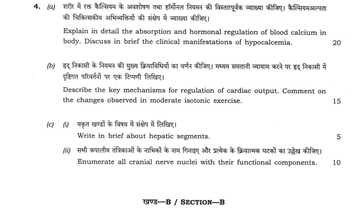
(a) Explain in detail the absorption and hormonal regulation of blood calcium in body. Discuss in brief the clinical…
Explain in detail the absorption and hormonal regulation of blood calcium in body. Discuss in brief the clinical manifestations of hypocalcemia. 20 marks
Describe the key mechanisms for regulation of cardiac output. Comment on the changes observed in moderate isotonic exercise. 15 marks
Write in brief about hepatic segments. 5 marks
Enumerate all cranial nerve nuclei with their functional components. 10 marks
हिंदी में प्रश्न पढ़ें
शरीर में रक्त कैल्सियम के अवशोषण तथा हार्मोनल नियमन की विस्तारपूर्वक व्याख्या कीजिए। कैल्सियमअल्पता की चिकित्सकीय अभिव्यक्तियों की संक्षेप में व्याख्या कीजिए। 20
हृद निकासी के नियमन की मुख्य क्रियाविधियों का वर्णन कीजिए। मध्यम समतानी व्यायाम करने पर हृद निकासी में दृष्टिगत परिवर्तनों पर एक टिप्पणी लिखिए। 15
यकृत खंडों के विषय में संक्षेप में लिखिए। 5
सभी कपालीय तंत्रिकाओं के नाभिकों के नाम गिनाइए और प्रत्येक के क्रियात्मक घटकों का उल्लेख कीजिए। 10
Model answer
Written by UPSC Answer Check against this question's marking rubric, to the expected length. UPSC does not publish answers for Mains — this is one way to score well, not an official key.
Calcium homeostasis and hypocalcaemia. Blood calcium is kept near 2.2–2.6 mmol/L by intestinal absorption, bone exchange and renal handling. Ionised calcium is sensed by parathyroid and renal calcium-sensing receptors; a fall triggers PTH release, while a rise suppresses PTH and favours calcitonin. In the skin, 7-dehydrocholesterol is converted to cholecalciferol by UVB; the liver hydroxylates it to 25-hydroxyvitamin D3, and the kidney, under PTH, converts it to 1,25-dihydroxyvitamin D3. This active vitamin D3 increases intestinal calcium absorption by upregulating TRPV6 channels, calbindin-D9K for cytoplasmic transport, and basolateral Ca2+-ATPase/Na+/Ca2+ exchangers. PTH is the principal hypercalcaemic hormone: it stimulates osteoclast-mediated bone resorption indirectly through osteoblast RANKL/OPG signalling, increases renal proximal and distal tubular calcium reabsorption, and increases renal 1α-hydroxylase activity. Calcitonin, from thyroid C cells, antagonises PTH by inhibiting osteoclast activity and reducing bone resorption, thereby lowering calcium. In the Indian subcontinent, vitamin D deficiency is common because of low sunlight exposure, dietary insufficiency and malnutrition, so hypocalcaemia is clinically important in malnourished children and elderly Indians. Manifestations reflect neuromuscular irritability: perioral and acral paraesthesia, carpopedal spasm, laryngospasm, seizures, Chvostek’s sign (facial nerve twitch on tapping) and Trousseau’s sign (carpal spasm with cuff inflation); ECG shows QT prolongation.
Cardiac output regulation. Cardiac output (CO = HR × stroke volume) is regulated by preload, afterload, contractility and autonomic tone. The Frank-Starling mechanism matches output to venous return: greater end-diastolic volume increases sarcomere length and force of contraction. Sympathetic β1 stimulation increases heart rate, conduction and contractility, while vagal withdrawal removes parasympathetic restraint on the SA node. Metabolic factors, circulating catecholamines and intracellular calcium enhance contractility. During moderate isotonic exercise, skeletal-muscle pump and respiratory pump increase venous return, HR rises, stroke volume is maintained or slightly increased, and CO rises about two- to three-fold, e.g. from 5 to 10–15 L/min. Sympathetic vasoconstriction in splanchnic and renal beds redistributes blood to active muscle, while local vasodilation in exercising muscle lowers peripheral resistance. On a cardiac output curve, the operating point shifts rightward with increased venous return and sympathetic drive, while afterload in non-exercising beds is maintained.
Hepatic segments and cranial nerve nuclei. Couinaud’s classification divides the liver into eight functionally independent segments by portal-vein branching and hepatic-vein drainage. Each segment contains its own portal triad (portal vein, hepatic artery, bile duct) and drains via hepatic veins: I caudate, II left lateral superior, III left lateral inferior, IV left medial, V right anterior inferior, VI right posterior inferior, VII right posterior superior and VIII right anterior superior. Segmental independence means one segment can be resected while preserving inflow/outflow of others, permitting segmental resection in hepatocellular carcinoma at Indian tertiary centres. Cranial nerve nuclei are best enumerated by functional columns: GSE, GVE, SVE, SVA, GVA and SSA. CN I olfactory bulb (SVA); CN II optic tract and lateral geniculate nucleus (SSA); CN III oculomotor nucleus (GSE) and Edinger-Westphal nucleus (GVE); CN IV trochlear nucleus (GSE); CN V motor nucleus (SVE), principal sensory and spinal trigeminal nuclei (GVA), mesencephalic nucleus (SSA); CN VI abducens nucleus (GSE); CN VII facial nucleus (GSE), superior salivatory nucleus (GVE), nucleus solitarius (SVA); CN VIII cochlear and vestibular nuclei (SSA); CN IX nucleus solitarius (SVA and GVA), nucleus ambiguus (SVE), inferior salivatory nucleus (GVE); CN X dorsal motor nucleus of vagus (GVE), nucleus ambiguus (SVE; also counted as GVE/parasympathetic for X), nucleus solitarius (SVA and GVA); CN XI nucleus ambiguus for cranial root (SVE) and spinal accessory nucleus (GSE of spinal root); CN XII hypoglossal nucleus (GSE). Thus calcium, circulation and neuroanatomy are integrated: hormonal control prevents tetany, cardiovascular regulation sustains perfusion, and segmental/cranial anatomy guides surgical and clinical assessment.
What "Explain" is asking you to do
Make the working of something clear — what sets it off, what follows from what, and what it produces. Explain is the Commission's mechanism word: it dominates the technical papers and the “explain why” stems, where the marks sit in the causal chain and not in the label.
Structure that answers it
State what it is → the initiating condition → the chain of cause, step by step → an instance where it plays out → what the chain produces
Where marks are lost
Describing what something looks like instead of why it works that way. Naming the stages without linking them reads as description too.
How this answer will be evaluated
Approach
Framework: Clinical Sequence (Definition > Aetiology/Pathophysiology > Features > Investigation/Management). (a) explain: definition/context > points in order > small example > short close | (b) describe: define > structure or process in order > labelled diagram > significance | (c(i)) write short notes: define > 3-4 key features > one example > one-line significance | (c(ii)) enumerate: list the items in order > one line each > no commentary Full marks: Comprehensive, accurate, and well-structured answers with all required mechanisms and clinical details.
Key points expected
- Intestinal absorption via Vitamin D (1,25-dihydroxycholecalciferol)
- Parathyroid hormone (PTH) action on bone and kidney
- Calcitonin role in calcium lowering
- Clinical manifestations of hypocalcemia (e.g., tetany, Chvostek's sign)
- Definition of Cardiac Output (CO = HR x SV)
- Mechanisms: Heart rate, Stroke volume, Preload, Afterload
- Changes in moderate isotonic exercise (increased venous return)
- Frank-Starling mechanism application
Evaluation rubric
Each sub-part is marked on its own, against the marks and word limit printed on the paper.
- (a) Detailed mechanism of calcium absorption and hormonal regulation, plus clinical features of hypocalcemia. 20 marks
explain— definition/context → points in order → small example → short close
Must cover
- Intestinal absorption via Vitamin D (1,25-dihydroxycholecalciferol)
- Parathyroid hormone (PTH) action on bone and kidney
- Calcitonin role in calcium lowering
- Clinical manifestations of hypocalcemia (e.g., tetany, Chvostek's sign)
Loses marks
- Listing symptoms without explaining the mechanism
- Omitting the role of Vitamin D
- Confusing hypercalcemia with hypocalcemia features
Earns more
- Mention of renal reabsorption mechanisms
- Reference to Vitamin D synthesis pathway
- Differentiation of acute vs chronic hypocalcemia
Extra mark
- Mention of specific serum calcium levels
- Reference to ECG changes in hypocalcemia
- (b) Mechanisms of cardiac output regulation and specific changes during moderate isotonic exercise. 15 marks
describe— define → structure or process in order → labelled diagram → significance
Must cover
- Definition of Cardiac Output (CO = HR x SV)
- Mechanisms: Heart rate, Stroke volume, Preload, Afterload
- Changes in moderate isotonic exercise (increased venous return)
- Frank-Starling mechanism application
Loses marks
- Failing to distinguish isotonic from isometric changes
- Ignoring the role of venous return
- Vague description of mechanisms
Earns more
- Mention of sympathetic nervous system role
- Distinction between isotonic and isometric exercise
- Reference to venous return and muscle pump
Extra mark
- Graphical representation of CO changes
- Mention of specific heart rate values
- (c(i)) Brief overview of hepatic segments. 5 marks
write short notes— define → 3-4 key features → one example → one-line significance
Must cover
- Definition of hepatic segments
- Couinaud classification (8 segments)
- Basis of segmentation (portal vein/vein of liver)
- Clinical significance (surgical resection)
Loses marks
- Confusing lobes with segments
- Omitting the basis of segmentation
- Vague description without specific names
Earns more
- Mention of functional vs anatomical segments
- Reference to hepatic veins as boundaries
Extra mark
- Diagram of Couinaud segments
- Mention of specific segmental resections
- (c(ii)) List of all cranial nerve nuclei with their functional components. 10 marks
enumerate— list the items in order → one line each → no commentary
Must cover
- List of all 12 cranial nerves
- Identification of nuclei for each nerve
- Functional components (Somatic, Special Somatic, General Visceral, Special Visceral)
- Correct association of nuclei with specific nerves
Loses marks
- Omitting functional components
- Incorrect association of nuclei
- Listing nerves without nuclei
Earns more
- Mention of specific nuclei names (e.g., Dorsal Motor Nucleus of Vagus)
- Clear tabular format
- Distinction between general and special visceral
Extra mark
- Mention of specific functional tests
- Reference to clinical syndromes
Practice this exact question
Write your answer and it is marked point by point against the model answer above — what you covered, what you missed, what you got wrong.
Evaluate my answer →More from Medical Science 2021 Paper I
- Q1 (a) Describe the formation, course, tributaries and termination of coronary sinus. 10 mar…
- Q2 (a) Describe the prostate under the following headings: (i) Gross features (ii) Lobes (ii…
- Q3 (a) Elaborate on the hormonal regulation of blood glucose level. Explain in brief the sig…
- Q4 (a) Explain in detail the absorption and hormonal regulation of blood calcium in body. Di…
- Q5 (a) Define 'metastasis'. Enumerate the steps involved in metastasis. Write briefly about…
- Q6 (a) Define ischemic heart disease. Enumerate four ischemic syndromes. Discuss the pathoge…
- Q7 (a) Define poison and classify it. Describe the treatment of venomous snakebite. Write a…