Paper II — Q3
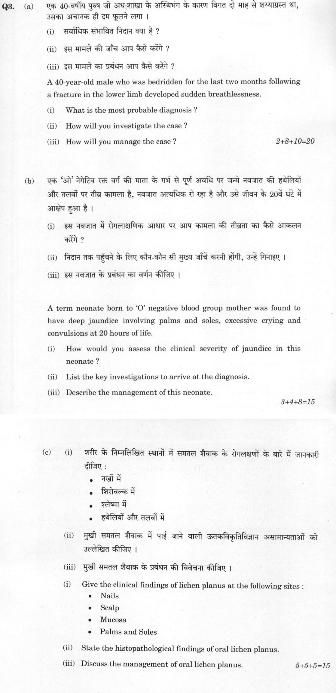
(a) A 40-year-old male who was bedridden for the last two months following a fracture in the lower limb developed sudden…
A 40-year-old male who was bedridden for the last two months following a fracture in the lower limb developed sudden breathlessness. What is the most probable diagnosis ?
How will you investigate the case ?
How will you manage the case ? (2+8+10=20 marks)
A term neonate born to 'O' negative blood group mother was found to have deep jaundice involving palms and soles, excessive crying and convulsions at 20 hours of life. How would you assess the clinical severity of jaundice in this neonate ?
List the key investigations to arrive at the diagnosis.
Describe the management of this neonate. (3+4+8=15 marks)
Give the clinical findings of lichen planus at the following sites : - Nails - Scalp - Mucosa - Palms and Soles
State the histopathological findings of oral lichen planus.
Discuss the management of oral lichen planus. (5+5+5=15 marks)
हिंदी में प्रश्न पढ़ें
एक 40-वर्षीय पुरुष जो अधःशाखा के अस्थिभंग के कारण विगत दो माह से शय्याप्रस्त था, उसका अचानक ही दम फूलने लगा । सर्वाधिक संभावित निदान क्या है ?
इस मामले की जाँच आप कैसे करेंगे ?
इस मामले का प्रबंधन आप कैसे करेंगे ? 2+8+10=20
एक 'ओ' नैगेटिव रक्त वर्ग की माता के गर्भ से पूर्ण अवधि पर जन्मे नवजात की हथेलियों और तलवों पर तीव्र कामला है, नवजात अत्यधिक रो रहा है और उसे जीवन के 20वें घंटे में आक्षेप हुआ है । इस नवजात में रोगलाक्षणिक आधार पर आप कामला की तीव्रता का कैसे आकलन करेंगे ?
निदान तक पहुँचने के लिए कौन-कौन सी मुख्य जाँचें करनी होंगी, उन्हें गिनाइए ।
इस नवजात के प्रबंधन का वर्णन कीजिए । 3+4+8=15
शरीर के निम्नलिखित स्थानों में समतल शेवाक के रोगलक्षणों के बारे में जानकारी दीजिए : - नखों में - शिरोवल्क में - श्लेष्मा में - हथेलियों और तलवों में
मुखी समतल शेवाक में पाई जाने वाली उतकविकृतिविज्ञान असामान्यताओं को उल्लेखित कीजिए ।
मुखी समतल शेवाक के प्रबंधन की विवेचना कीजिए । 5+5+5=15
Model answer
Written by UPSC Answer Check against this question's marking rubric, to the expected length. UPSC does not publish answers for Mains — this is one way to score well, not an official key.
Section (a): Pulmonary Embolism
Probable Diagnosis: The most probable diagnosis is Acute Pulmonary Embolism (PE) secondary to deep vein thrombosis (DVT). The patient fits Virchow's triad due to stasis from prolonged immobilization following a lower limb fracture, endothelial injury, and hypercoagulability.
Investigations: Initial clinical pre-test probability is calculated using the Wells score or revised Geneva score.
- Bedside tests: ECG classically shows sinus tachycardia, or the S_1Q_3T₃ pattern (deep S wave in I, Q wave and inverted T wave in III), along with right bundle branch block and right ventricular strain. Arterial Blood Gas (ABG) typically demonstrates hypoxemia, hypocapnia, and respiratory alkalosis.
- Blood investigations: Quantitative D-dimer (ELISA) has a high negative predictive value to rule out PE in low-to-moderate risk cases. Elevated cardiac biomarkers (Troponin I/T and NT-proBNP) assess right ventricular (RV) microinfarction and strain.
- Imaging: CT Pulmonary Angiography (CTPA) is the definitive gold standard imaging modality. Transthoracic Echocardiography (TTE) identifies RV dilatation, RV dysfunction, and McConnell’s sign. Compression Duplex Ultrasonography of lower limbs evaluates the source DVT. Ventilation-Perfusion (V/Q) scan is indicated if CTPA is contraindicated (e.g., severe renal impairment).
Management:
- Resuscitation: High-flow oxygen supplementation to maintain saturation >90%, intravenous crystalloid fluids (cautiously to avoid RV overload), and vasopressor support (Norepinephrine) if hemodynamically unstable.
- Systemic Thrombolysis: Indicated for massive PE (hemodynamically unstable with persistent hypotension or shock). Intravenous recombinant tissue plasminogen activator (rtPA/Alteplase 100 mg over 2 hours) or Tenecteplase is administered. Surgical or catheter-directed embolectomy is considered if thrombolysis is contraindicated.
- Anticoagulation: For hemodynamically stable patients, therapeutic anticoagulation is initiated immediately using Low Molecular Weight Heparin (LMWH, e.g., Enoxaparin 1 mg/kg subcutaneous twice daily), Fondaparinux, or unfractionated heparin, bridged to Warfarin (target INR 2.0–3.0) or transitioned to Direct Oral Anticoagulants (DOACs like Rivaroxaban or Apixaban) for a minimum of 3 to 6 months.
- IVC Filter: An Inferior Vena Cava (IVC) filter is placed if anticoagulation is strictly contraindicated or in recurrent PE despite therapeutic anticoagulation.
---
Section (b): Neonatal Hemolytic Jaundice
Assessment of Clinical Severity: The onset of jaundice at <24 hours of life involving the palms and soles indicates severe pathological hyperbilirubinemia, staged clinically by Kramer’s Rule (Zone 5 involvement). Neurological symptoms like excessive crying (high-pitched cry) and convulsions signify Acute Bilirubin Encephalopathy (Kernicterus risk) due to free unconjugated bilirubin crossing the blood-brain barrier.
Key Investigations:
- Total and conjugated/unconjugated serum bilirubin levels.
- Blood grouping and Rh typing of infant and mother to confirm Rh/ABO isoimmunization.
- Direct Antiglobulin Test (Direct Coombs Test) to detect maternal antibodies on neonatal RBCs; maternal indirect Coombs test for antibody titres.
- Peripheral blood smear (revealing spherocytes or reticulocytosis) and Reticulocyte count (elevated, indicating hemolysis).
- Complete blood count for hemoglobin/hematocrit and serum albumin.
Management:
- Immediate Intensive Phototherapy: Continuous, high-intensity narrow-band blue light (460--490 nm) placed within30 cm of the infant to maximize skin exposure, converting unconjugated bilirubin into water-soluble lumirubin.
- Double Volume Exchange Transfusion (DVET): Indicated urgently due to encephalopathic signs (convulsions) and critically elevated bilirubin levels crossing exchange thresholds on AAP nomograms. It removes sensitized RBCs, circulating antibodies, and free bilirubin using compatible reconstituted blood.
- Intravenous Immunoglobulin (IVIG): High-dose IVIG (0.5--1 g/kg over 2 hours) is administered to block Fc receptors on reticuloendothelial cells and arrest ongoing hemolysis.
- Supportive Care and Neurological Monitoring: Control convulsions with phenobarbitone, ensure hydration, correct acidosis, and follow up with Brainstem Evoked Response Audiometry (BERA) for neurodevelopmental sequelae.
---
Section (c): Lichen Planus
Site-Specific Clinical Findings:
- Nails: Thinning of the nail plate, longitudinal ridging, grooving, subungual hyperkeratosis, and pathognomonic dorsal pterygium formation leading to permanent anonychia.
- Scalp: Lichen planopilaris presenting as follicular hyperkeratotic papules, peripilar erythema, and end-stage scarring/cicatricial alopecia.
- Mucosa: Oral cavity displays Wickham’s striae (lacy, white reticular lines, especially on the buccal mucosa), along with erosive, atrophic, or bullous lesions causing severe burning.
- Palms and Soles: Palmoplantar lichen planus presents as compact, yellowish, hyperkeratotic plaques with central punctate pitting or erosions, often mimicking callosities or hypertrophic variants.
Histopathology of Oral Lichen Planus: Characteristic microscopic hallmarks include:
- Hyperkeratosis with liquefaction degeneration of the basal cell layer.
- "Saw-tooth" appearance of the rete ridges.
- Dense, band-like (lichenoid) lymphocytic infiltrate strictly confined to the upper dermis/lamina propria.
- Civatte bodies (colloid/apoptotic bodies) representing degenerated basal keratinocytes.
- Cleft formation between the epithelium and connective tissue known as Max-Joseph spaces.
Management of Oral Lichen Planus:
- First-line therapy: High-potency topical corticosteroids (Clobetasol propionate 0.05% gel, Triamcinolone acetonide 0.1% paste) applied directly to lesions.
- Second-line therapy: Topical calcineurin inhibitors (Tacrolimus 0.1% or Pimecrolimus ointment) for resistant mucosal lesions.
- Systemic agents: Short courses of systemic oral corticosteroids (Prednisolone) for severe exacerbations; systemic retinoids (Acitretin) or immunosuppressants (Methotrexate, Azathioprine, Mycophenolate Mofetil) in refractory disease.
- Surveillance: Elimination of local irritants, maintenance of oral hygiene, and regular long-term follow-up to monitor for malignant transformation into oral squamous cell carcinoma.
What "Describe" is asking you to do
Give a full, ordered account of the thing named — its parts, stages or mechanism — in the sequence in which it actually exists or occurs. Most describe questions come from the science optionals, where the marks sit in correct technical detail and, where the stem says so, a labelled diagram.
Structure that answers it
One-line identification of the subject → the parts or stages in their real order, each with its defining detail → labelled diagram where the subject is structural → closing line on function or significance
Where marks are lost
Loose general prose where the examiner is ticking named parts, correct terminology and their sequence; and in the General Studies papers, turning to evaluation before the description is finished.
How this answer will be evaluated
Approach
Framework: Clinical Sequence (Diagnosis > Investigation > Management). (a(i)) explain: definition/context > points in order > small example > short close | (a(ii)) explain: definition/context > points in order > small example > short close | (a(iii)) explain: definition/context > points in order > small example > short close | (b(i)) explain: definition/context > points in order > small example > short close | (b(ii)) enumerate: list the items in order > one line each > no commentary | (b(iii)) explain: definition/context > points in order > small example > short close | (c(i)) describe: define > structure or process in order > labelled diagram > significance | (c(ii)) explain: definition/context > points in order > small example > short close | (c(iii)) explain: definition/context > points in order > small example > short close Full marks: Accurate diagnosis, prioritized investigations, tiered management, specific clinical/histological details.
Key points expected
- Pulmonary Embolism (PE)
- Mention risk factor: bedridden/immobility
- Mention risk factor: lower limb fracture
- D-dimer (screening)
- CT Pulmonary Angiography (CTPA) as gold standard
- ECG (S1Q3T3, RBBB, sinus tach)
- Chest X-ray (often normal or wedge-shaped infarct)
- Immediate: Oxygen, IV fluids, hemodynamic support
Evaluation rubric
Each sub-part is marked on its own, against the marks and word limit printed on the paper.
- (a(i)) Identify the most probable diagnosis based on the clinical vignette. 2 marks
explain— definition/context → points in order → small example → short close
Must cover
- Pulmonary Embolism (PE)
- Mention risk factor: bedridden/immobility
- Mention risk factor: lower limb fracture
Loses marks
- Diagnosing Pneumonia or Heart Failure without PE
- Failing to link diagnosis to the specific risk factors
Earns more
- Mention DVT as precursor
- (a(ii)) List and explain the investigation protocol for the case. 8 marks
explain— definition/context → points in order → small example → short close
Must cover
- D-dimer (screening)
- CT Pulmonary Angiography (CTPA) as gold standard
- ECG (S1Q3T3, RBBB, sinus tach)
- Chest X-ray (often normal or wedge-shaped infarct)
Loses marks
- Listing investigations without priority (e.g., CTPA before D-dimer)
- Omitting the gold standard diagnostic test
Earns more
- Mentioning V/Q scan if CTPA contraindicated
- Mentioning Echocardiogram for RV strain
- Mentioning ABG for hypoxemia
Extra mark
- Mentioning Wells Score for pre-test probability
- (a(iii)) Describe the management plan in tiers (immediate, definitive, follow-up). 10 marks
explain— definition/context → points in order → small example → short close
Must cover
- Immediate: Oxygen, IV fluids, hemodynamic support
- Definitive: Anticoagulation (Heparin followed by Warfarin/DOACs)
- Thrombolysis (tPA) if hemodynamically unstable
- Follow-up: IVC filter if anticoagulation contraindicated
Loses marks
- Management without priority (e.g., starting oral meds before IV heparin)
- Omitting the role of thrombolysis in massive PE
Earns more
- Mentioning duration of anticoagulation (3-6 months)
- Mentioning monitoring of INR if using Warfarin
Extra mark
- Mentioning specific guidelines (e.g., ESC or AHA guidelines)
- (b(i)) Assess the clinical severity of jaundice in the neonate. 3 marks
explain— definition/context → points in order → small example → short close
Must cover
- Kramer's zones (palms/soles = Zone 5)
- Mentioning 'deep jaundice' implies high bilirubin
- Mentioning neurological signs (convulsions) as severity marker
Loses marks
- Assessing severity only by color without zones
- Ignoring the neurological symptoms in the assessment
Earns more
- Mentioning 'Acute Bilirubin Encephalopathy' (ABE) signs
- Mentioning the timing (20 hours) as a risk factor
- (b(ii)) List key investigations to arrive at the diagnosis. 4 marks
enumerate— list the items in order → one line each → no commentary
Must cover
- Total and Direct Bilirubin levels
- Blood grouping (Mother and Child)
- Coombs test (Direct and Indirect)
- Peripheral blood smear (for hemolysis)
Loses marks
- Omitting the Coombs test
- Omitting blood grouping of both mother and child
Earns more
- Mentioning Reticulocyte count
- Mentioning G6PD deficiency test (if relevant)
- (b(iii)) Describe the management of this neonate. 8 marks
explain— definition/context → points in order → small example → short close
Must cover
- Immediate: IV fluids and glucose
- Definitive: Phototherapy (Intensive)
- Definitive: Exchange transfusion (due to convulsions/ABE)
- Follow-up: Anti-D immunoglobulin for mother
Loses marks
- Management without priority (e.g., phototherapy only for ABE)
- Omitting the need for exchange transfusion in severe cases
Earns more
- Mentioning specific criteria for exchange transfusion
- Mentioning monitoring of bilirubin levels
Extra mark
- Mentioning specific national guidelines for neonatal jaundice
- (c(i)) Give clinical findings of lichen planus at specific sites. 5 marks
describe— define → structure or process in order → labelled diagram → significance
Must cover
- Nails: Longitudinal ridging, pterygium, thinning
- Scalp: Scarring alopecia (frontal fibrosing alopecia)
- Mucosa: Wickham's striae, erosions, atrophy
- Palms/Soles: Hyperkeratotic papules, pits
Loses marks
- Describing only one site in detail and ignoring others
- Confusing lichen planus with lichen simplex chronicus
Earns more
- Mentioning 'Wickham's striae' specifically for mucosa
- Mentioning 'scarring' for scalp involvement
- (c(ii)) State the histopathological findings of oral lichen planus. 5 marks
explain— definition/context → points in order → small example → short close
Must cover
- Hyperkeratosis (orthokeratosis or parakeratosis)
- Saw-tooth appearance of rete ridges
- Band-like lymphocytic infiltrate at dermo-epidermal junction
- Civatte bodies (apoptotic keratinocytes)
Loses marks
- Omitting the 'saw-tooth' rete ridges
- Omitting the 'band-like' nature of the infiltrate
Earns more
- Mentioning 'basal cell degeneration'
- Mentioning 'liquefaction degeneration' of basal layer
- (c(iii)) Discuss the management of oral lichen planus. 5 marks
explain— definition/context → points in order → small example → short close
Must cover
- Immediate: Topical corticosteroids (first line)
- Definitive: Systemic steroids (for severe/refractory cases)
- Follow-up: Regular monitoring for malignant transformation
- Mentioning 'avoidance of irritants' (spicy food, alcohol)
Loses marks
- Management without priority (e.g., systemic steroids first)
- Omitting the need for long-term monitoring
Earns more
- Mentioning 'calcineurin inhibitors' as an alternative
- Mentioning 'psychological support' for chronic condition
Extra mark
- Mentioning specific long-term follow-up intervals (e.g., every 6 months)
Practice this exact question
Write your answer and it is marked point by point against the model answer above — what you covered, what you missed, what you got wrong.
Evaluate my answer →More from Medical Science 2021 Paper II
- Q1 (a) Discuss in brief the role of ultrasound in the investigation of pain in right hypocho…
- Q2 (a) A sixty-year-old diabetic male presented to the emergency department with acute onset…
- Q3 (a) A 40-year-old male who was bedridden for the last two months following a fracture in…
- Q4 (a) Discuss in short the etiology, diagnosis, treatment and complications of cirrhosis of…
- Q5 (a) Briefly describe the clinical features, diagnostic workup and management of a patient…
- Q6 (a) (i) Write in brief the reasons for rising Caesarean Section rates globally and in Ind…