Paper I — Q1
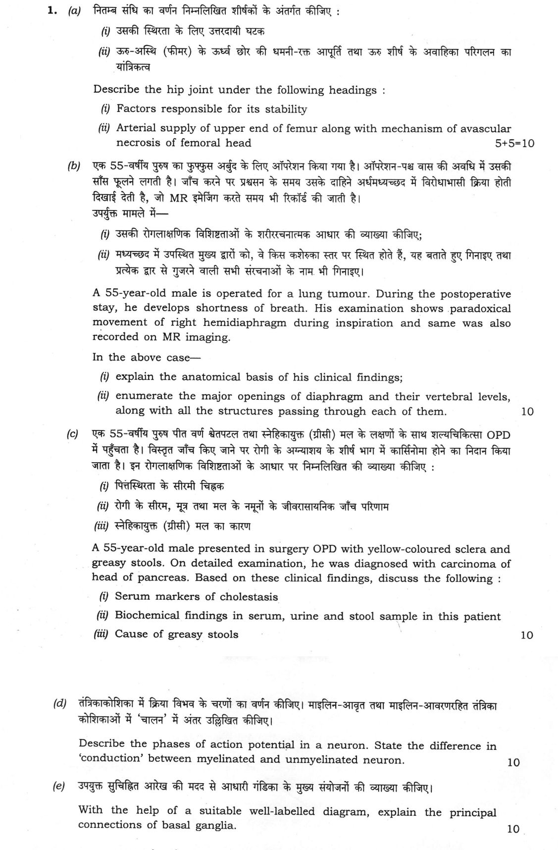
(a) Describe the hip joint under the following headings : (i) Factors responsible for its stability (ii) Arterial supply of upper…
Describe the hip joint under the following headings : Factors responsible for its stability
Arterial supply of upper end of femur along with mechanism of avascular necrosis of femoral head 10 marks
A 55-year-old male is operated for a lung tumour. During the postoperative stay, he develops shortness of breath. His examination shows paradoxical movement of right hemidiaphragm during inspiration and same was also recorded on MR imaging. In the above case— explain the anatomical basis of his clinical findings;
enumerate the major openings of diaphragm and their vertebral levels, along with all the structures passing through each of them. 10 marks
A 55-year-old male presented in surgery OPD with yellow-coloured sclera and greasy stools. On detailed examination, he was diagnosed with carcinoma of head of pancreas. Based on these clinical findings, discuss the following : Serum markers of cholestasis
Biochemical findings in serum, urine and stool sample in this patient
Cause of greasy stools 10 marks
Describe the phases of action potential in a neuron. State the difference in ‘conduction’ between myelinated and unmyelinated neuron. 10 marks
With the help of a suitable well-labelled diagram, explain the principal connections of basal ganglia. 10 marks
हिंदी में प्रश्न पढ़ें
निम्न सन्धि का वर्णन निम्नलिखित शीर्षकों के अंतर्गत कीजिए : उसकी स्थिरता के लिए उत्तरदायी घटक
ऊरु-अस्थि (फीमर) के ऊर्ध्व छोर की धमनी-रक्त आपूर्ति तथा ऊरु शीर्ष के अवाहिका परिगलन का यांत्रिकत्व (10 अंक)
एक 55-वर्षीय पुरुष का फुफ्फुस अर्बुद के लिए ऑपरेशन किया गया है। ऑपरेशन-पश्च वास की अवधि में उसकी साँस फूलने लगती है। जाँच करने पर प्रश्वसन के समय उसके दाहिने अर्धमध्यच्छद में विरोधाभासी क्रिया होती दिखाई देती है, जो MR इमेजिंग करते समय भी रिकॉर्ड की जाती है। उपर्युक्त मामले में— उसकी रोगलाक्षणिक विशिष्टताओं के शारीरचनात्मक आधार की व्याख्या कीजिए;
मध्यच्छद में उपस्थित मुख्य द्वारों को, वे किस कशेरुका स्तर पर स्थित होते हैं, यह बताते हुए गिनाइए तथा प्रत्येक द्वार से गुजरने वाली सभी संरचनाओं के नाम भी गिनाइए। (10 अंक)
एक 55-वर्षीय पुरुष पीत वर्ण श्वेतपटल तथा स्नेहिकायुक्त (ग्रीसी) मल के लक्षणों के साथ शल्यचिकित्सा OPD में पहुँचता है। विस्तृत जाँच किए जाने पर रोगी के अग्न्याशय के शीर्ष भाग में कार्सिनोमा होने का निदान किया जाता है। इन रोगलाक्षणिक विशिष्टताओं के आधार पर निम्नलिखित की व्याख्या कीजिए : पित्तस्थिरता के सीरम चिह्नक
रोगी के सीरम, मूत्र तथा मल के नमूनों के जीवरासायनिक जाँच परिणाम
स्नेहिकायुक्त (ग्रीसी) मल का कारण (10 अंक)
तंत्रिकाकोशिका में क्रिया विभव के चरणों का वर्णन कीजिए। माइलिन-आवृत तथा माइलिन-आवरणरहित तंत्रिका कोशिकाओं में ‘चालन’ में अंतर उल्लिखित कीजिए। (10 अंक)
उपयुक्त सुचिह्नित आरेख की मदद से आधारी गंडिका के मुख्य संयोजनों की व्याख्या कीजिए। (10 अंक)
Model answer
Drafted with AI by UPSC Answer Check against this question's marking rubric, to the expected length. UPSC does not publish answers for Mains — this is one way to score well, not an official key. How we build rubrics and answers.
(a)(i) Factors responsible for the stability of the hip joint (5 marks)
The hip is a ball-and-socket synovial joint, and it is the most stable large joint in the body because of its bony, ligamentous and muscular arrangements.
- Bony: the femoral head (about two-thirds of a sphere) sits deeply in the acetabulum, which is deepened by the fibrocartilaginous labrum that grips the head beyond its equator. The transverse acetabular ligament closes the acetabular notch.
- Capsule and ligaments: the strong fibrous capsule is reinforced by three ligaments that twist and tighten in extension. The iliofemoral (Y-shaped ligament of Bigelow) is the strongest ligament in the body and prevents hyperextension and holds the trunk erect in standing, the pubofemoral limits abduction and extension, and the ischiofemoral limits extension and medial rotation. The ligament of the head of the femur is only a minor stabiliser.
- Atmospheric pressure and synovial fluid: the negative intra-articular pressure and the cohesion of the fluid film form a suction seal.
- Muscles: the surrounding muscles give dynamic stability, especially gluteus medius and minimus, which hold the pelvis level in single-leg stance, with the short lateral rotators posteriorly and iliopsoas anteriorly.
(a)(ii) Arterial supply of the upper end of the femur and avascular necrosis of the head (5 marks)
Arterial supply. The upper end of the femur has three sources.
- The retinacular arteries from the trochanteric (cruciate) anastomosis, formed by the medial and lateral circumflex femoral arteries (branches of the profunda femoris) with the superior and inferior gluteal arteries. The medial circumflex femoral supplies the largest share, through the posterosuperior and posteroinferior retinacular branches. These run upwards along the neck, under the reflected synovium and within the capsule, to enter the head near the articular margin. The lateral circumflex supplies the anterior retinacular vessels.
- The artery of the ligament of the head (from the acetabular branch of the obturator artery), which supplies only a small area around the fovea and is inadequate in adults.
- The nutrient (metaphyseal) artery to the shaft, which reaches the neck only in part.
Mechanism of avascular necrosis. In the adult, the epiphyseal plate is closed and the metaphyseal supply does not reach the head. So the head depends mainly on the retinacular vessels. An intracapsular fracture of the neck (subcapital), or a dislocation, tears or kinks these vessels, or the vessels are occluded (by steroids, alcohol, sickle-cell disease, or decompression). The result is:
- Ischaemia and death of bone and marrow in the head, with the articular cartilage surviving for a time on synovial fluid.
- Weak, revascularising bone, since new bone is laid down on dead trabeculae (creeping substitution), which then collapses under load, giving a subchondral fracture (crescent sign) and flattening of the head.
- Secondary osteoarthritis, pain and limitation of movement.
(b) Paradoxical right hemidiaphragm after lung surgery (10 marks)
(i) Anatomical basis. The right phrenic nerve (C3, C4, C5, mainly C4) supplies the motor and sensory innervation of the right hemidiaphragm. In the thorax it runs down on the right side of the mediastinum along the superior vena cava and right atrium, in front of the root of the lung, over the pericardium. In this patient it has probably been injured during hilar and mediastinal dissection for the lung tumour (or by tumour infiltration). The right dome is then paralysed. In a normal inspiration, the left dome contracts and descends, creating negative intrathoracic pressure, and the paralysed dome, being flaccid, is passively pulled upwards into the chest, so it moves paradoxically, up on inspiration and down on expiration. This gives less effective ventilation, basal collapse and shortness of breath, and it shows on a sniff test on fluoroscopy, ultrasound and dynamic MR imaging.
(ii) Openings of the diaphragm.
- Caval opening, T8 (in the central tendon, to the right of the midline): the inferior vena cava and the branches of the right phrenic nerve.
- Oesophageal opening, T10 (in the muscle of the right crus): the oesophagus, the anterior and posterior vagal trunks, the oesophageal branches of the left gastric vessels, and lymphatics.
- Aortic opening, T12 (behind the median arcuate ligament): the aorta, the thoracic duct, and often the azygos vein.
- Other openings: the greater, lesser and least splanchnic nerves pierce the crura; the sympathetic trunk passes behind the medial arcuate ligament (with psoas); the subcostal nerve and vessels pass behind the lateral arcuate ligament (with quadratus lumborum); the hemiazygos vein pierces the left crus; and the superior epigastric vessels and lymphatics pass between the sternal and costal slips (foramen of Morgagni). The left phrenic nerve pierces the left dome of the muscle.
(c) Carcinoma of the head of the pancreas with obstructive jaundice (10 marks)
The tumour compresses the common bile duct where it runs through the pancreatic head, and it may also block the pancreatic duct. Bile cannot reach the gut, so the picture is that of post-hepatic (obstructive) jaundice with steatorrhoea.
(i) Serum markers of cholestasis.
- Raised conjugated (direct) bilirubin, so the jaundice is yellow-green.
- Markedly raised alkaline phosphatase (from the biliary canaliculi) and gamma-glutamyl transferase (GGT), with raised 5′-nucleotidase to show the hepatic origin of the ALP.
- Raised bile salts and cholesterol (lipoprotein X), and a mild rise in transaminases, which is a late and small change.
- Prolonged prothrombin time from vitamin K malabsorption, which is corrected by parenteral vitamin K. CA 19-9 is raised as a tumour marker.
(ii) Biochemical findings.
- Serum: conjugated hyperbilirubinaemia, with high ALP, GGT and bile salts, and raised cholesterol.
- Urine: bilirubin present (dark, frothy urine), since conjugated bilirubin is water-soluble and is filtered. Urobilinogen is absent or greatly reduced, since bile does not reach the gut where bacteria form it. Bile salts are present in the urine.
- Stool: pale, clay-coloured because of the absence of stercobilin, and bulky, offensive, and greasy with a high fat content. Stool urobilinogen (stercobilinogen) is absent.
(iii) Cause of the greasy stools (steatorrhoea). Fat is not digested and absorbed properly, for two reasons. Bile salts do not reach the duodenum, so fat is not emulsified and mixed micelles cannot form, and lipase action and absorption fall. If the pancreatic duct is blocked as well, pancreatic lipase and colipase are also missing, and fat stays undigested. The unabsorbed fat (more than 7 g/day) appears in the stool, which is pale, bulky, foul-smelling and floats. There is also malabsorption of the fat-soluble vitamins A, D, E and K.
(d) Action potential in a neuron and conduction in myelinated and unmyelinated fibres (10 marks)
The graph shows the phases. The resting membrane potential is about −70 mV, maintained by K+ leak channels and the Na+/K+ pump (3 Na+ out, 2 K+ in).

- Depolarisation: a stimulus opens a few voltage-gated Na+ channels. When the membrane reaches the threshold (about −55 mV), more open in a positive-feedback (Hodgkin) cycle, and the rapid Na+ influx drives the potential upwards.
- Overshoot: the potential reaches about +30 to +35 mV, near the Na+ equilibrium potential (about +60 mV).
- Repolarisation: the Na+ channels are inactivated (inactivation gate closes) and the voltage-gated K+ channels open, so K+ leaves the cell and the potential falls.
- After-hyperpolarisation: the K+ channels close slowly, and the potential dips towards the K+ equilibrium potential (about −80 to −90 mV), before it returns to rest through the pump and leak channels.
- Refractory periods: during the absolute refractory period, from depolarisation to about the middle of repolarisation, no stimulus can trigger a second spike since Na+ channels are inactivated; in the relative refractory period a stronger stimulus is needed. This makes the spike all-or-none and one-directional. The whole spike lasts 1–2 ms.
Conduction.
- Unmyelinated fibres: the impulse spreads continuously along the membrane, each patch depolarising the next through local currents. The velocity is slow (0.5–2 m/s), it rises with the square root of the fibre diameter, and the process uses much energy, since ions cross the whole membrane and the pump must restore them.
- Myelinated fibres: the myelin sheath insulates the axon and increases membrane resistance and lowers capacitance, and the voltage-gated Na+ channels are concentrated at the nodes of Ranvier (about 1 mm apart). The impulse jumps from node to node (saltatory conduction). Velocity is much higher (up to 120 m/s), rising in proportion to the fibre diameter, and less energy is used since only the nodes depolarise. Loss of myelin (multiple sclerosis, Guillain–Barré syndrome) slows or blocks conduction.
(e) Principal connections of the basal ganglia (10 marks)
The basal ganglia are the striatum (caudate nucleus and putamen), globus pallidus (external, GPe, and internal, GPi), the subthalamic nucleus (STN), and the substantia nigra (pars compacta, SNc, and pars reticulata, SNr). They modulate movement through loops with the cortex and the thalamus. The diagram shows the main connections.

Afferents. The striatum, the input nucleus, receives an excitatory glutamatergic projection from the whole cerebral cortex (corticostriatal, mainly the motor, premotor and prefrontal areas), from the intralaminar thalamic nuclei, and a dopaminergic projection from the substantia nigra pars compacta (nigrostriatal).
Efferents. The output nuclei, GPi and SNr, send inhibitory GABAergic fibres to the ventral anterior and ventral lateral nuclei of the thalamus, which project back to the premotor and motor cortex (the cortico-striato-pallido-thalamo-cortical loop), and to the brainstem (pedunculopontine nucleus and superior colliculus).
Pathways.
- Direct pathway: cortex excites striatal neurons that carry D1 receptors (GABA and substance P), which inhibit GPi and SNr. This releases the thalamus from inhibition, so the thalamus excites the cortex and movement is facilitated.
- Indirect pathway: cortex excites striatal neurons that carry D2 receptors (GABA and enkephalin), which inhibit GPe. GPe therefore stops inhibiting the STN, which excites GPi and SNr, and so the thalamus is inhibited more and movement is suppressed.
- Hyperdirect pathway: the cortex drives the STN directly, providing a fast brake.
- Dopamine from the SNc excites D1 neurons and inhibits D2 neurons, so both actions favour movement.
Clinical relevance. In Parkinson's disease the loss of nigral dopamine reduces the direct and enhances the indirect pathway, causing bradykinesia and rigidity. Degeneration of striatal neurons in Huntington's disease causes chorea, and a lesion of the STN causes hemiballismus.
How this answer will be evaluated
Evaluation rubric
Each sub-part is marked on its own, against the marks and word limit printed on the paper.
- (a) Hip joint: (i) factors responsible for stability; (ii) arterial supply of the upper end of the femur with the mechanism of avascular necrosis of the femoral head (5 + 5 = 10 marks). 10 marks
Must cover
- Stability: deep acetabulum, labrum and transverse ligament, iliofemoral, pubofemoral and ischiofemoral ligaments, capsule, suction and muscles
- Iliofemoral (Y) ligament as the strongest, preventing hyperextension
- Supply: retinacular arteries from medial and lateral circumflex femoral (trochanteric anastomosis), artery of ligament of head, nutrient artery
- Avascular necrosis: retinacular vessels torn in intracapsular fracture or dislocation, head left ischaemic, collapse and osteoarthritis
Loses marks
- Omitting the AVN mechanism.
- Stating that the ligament of the head is the main supply in adults.
- Confusing the hip with the shoulder.
Earns more
- Both (i) and (ii) answered under separate headings.
- Medial circumflex femoral artery identified as the main supplier.
Extra mark
- Causes such as steroids, alcohol and sickle cell disease; crescent sign
- (b) Post-thoracotomy paradoxical right hemidiaphragm: (i) anatomical basis; (ii) major diaphragmatic openings with vertebral levels and every structure through each (10 marks). 10 marks
Must cover
- Right phrenic nerve (C3–C5) injury during hilar or mediastinal dissection, with its course over the pericardium
- Paradoxical movement explained: paralysed dome rises on inspiration as the normal side descends and intrathoracic pressure falls
- Openings with levels: caval T8, oesophageal T10, aortic T12
- Contents of each opening plus the minor passages: sympathetic trunk, splanchnic nerves, subcostal vessels, superior epigastric vessels
Loses marks
- Wrong vertebral levels.
- Naming the vagus or the left phrenic nerve as the injured nerve.
- Listing the openings without their contents.
Earns more
- Contents correct for every opening: IVC and right phrenic branches; oesophagus and vagal trunks; aorta, thoracic duct and azygos vein.
- Mention of the clinical effects: dyspnoea, basal collapse, and the sniff test.
Extra mark
- Left phrenic nerve piercing the muscle; hemiazygos through the left crus
- (c) Carcinoma of the head of pancreas with jaundice and greasy stools: (i) serum markers of cholestasis; (ii) biochemical findings in serum, urine and stool; (iii) cause of greasy stools (10 marks). 10 marks
Must cover
- Serum: conjugated bilirubin, markedly raised ALP and GGT, raised bile salts and cholesterol
- Urine: bilirubin present, urobilinogen absent
- Stool: pale, clay-coloured with no stercobilin and increased fat
- Steatorrhoea from absent bile salts (no emulsification or micelles) and, if the duct is blocked, lack of pancreatic lipase
Loses marks
- Stating unconjugated bilirubin as the raised fraction.
- Urobilinogen raised in the urine.
- Leaving out the stool findings.
Earns more
- Findings given separately for serum, urine and stool.
- Reason given for each finding, including the water solubility of conjugated bilirubin.
Extra mark
- Prolonged prothrombin time, fat-soluble vitamin deficiency, CA 19-9
- (d) Phases of the neuronal action potential and the difference in conduction between myelinated and unmyelinated neurons (10 marks). 10 marks
Must cover
- Resting potential about −70 mV, threshold about −55 mV, and voltage-gated Na+ channel opening for depolarisation with overshoot to about +35 mV
- Repolarisation by Na+ inactivation and K+ efflux, then after-hyperpolarisation
- Absolute and relative refractory periods; all-or-none spike
- Myelinated: saltatory conduction at nodes of Ranvier, fast and energy-saving; unmyelinated: continuous, slow
Loses marks
- Describing K+ influx as the cause of depolarisation.
- Omitting the ionic basis of each phase.
- Reversing the conduction differences.
Earns more
- Labelled graph of potential against time.
- Typical velocities and dependence on fibre diameter.
Extra mark
- Effect of demyelination in multiple sclerosis or Guillain–Barré syndrome
- (e) Explain the principal connections of the basal ganglia with the help of a well-labelled diagram (10 marks). 10 marks
Must cover
- Components: striatum, GPe, GPi, STN and substantia nigra (SNc and SNr)
- Afferents: corticostriatal glutamatergic, nigrostriatal dopaminergic, thalamostriatal
- Efferents: GPi/SNr inhibit the VA/VL thalamus, which excites the motor and premotor cortex
- Direct pathway (facilitates), indirect pathway through GPe and STN (inhibits), and dopamine acting on D1 and D2 neurons
Loses marks
- Showing the thalamus as inhibited by the cortex.
- Omitting the STN or the substantia nigra.
- No diagram.
Earns more
- A labelled diagram showing excitatory and inhibitory arrows.
- Clinical correlation with Parkinson's disease, Huntington's disease or hemiballismus.
Extra mark
- Hyperdirect pathway
What "Describe" is asking you to do
Give a full, ordered account of the thing named — its parts, stages or mechanism — in the sequence in which it actually exists or occurs. Most describe questions come from the science optionals, where the marks sit in correct technical detail and, where the stem says so, a labelled diagram.
Structure that answers it
One-line identification of the subject → the parts or stages in their real order, each with its defining detail → labelled diagram where the subject is structural → closing line on function or significance
Where marks are lost
Loose general prose where the examiner is ticking named parts, correct terminology and their sequence; and in the General Studies papers, turning to evaluation before the description is finished.
Practice this exact question
Write your answer and it is marked point by point against the model answer above — what you covered, what you missed, what you got wrong.
Evaluate my answer →More from Medical Science 2026 Paper I
- Q1 (a) Describe the hip joint under the following headings : (i) Factors responsible for its…
- Q2 (a) A 32-year-old male is brought to the emergency department following a road traffic ac…
- Q3 (a) (i) 1. Name the ligaments of ovary and their role in ovarian torsion and its complica…
- Q4 (a) (i) Describe the cerebrospinal fluid (CSF) flow from site of production to its absorp…