Paper I — Q3
(a) (i) 1. Name the ligaments of ovary and their role in ovarian torsion and its complications. 2. Explain the surgical…
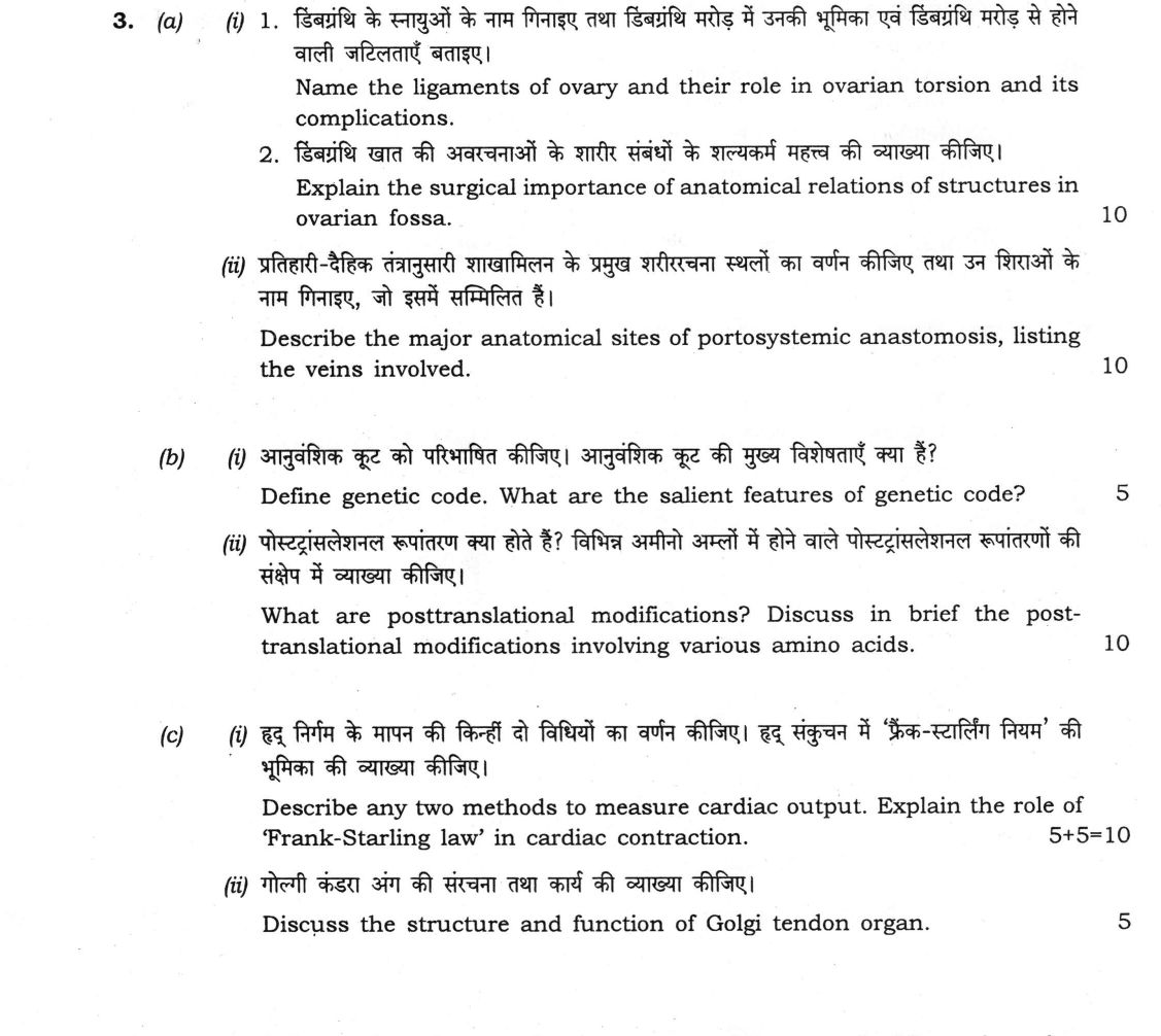
1. Name the ligaments of ovary and their role in ovarian torsion and its complications. 2. Explain the surgical importance of anatomical relations of structures in ovarian fossa. 10 marks
Describe the major anatomical sites of portosystemic anastomosis, listing the veins involved. 10 marks
Define genetic code. What are the salient features of genetic code? 5 marks
What are posttranslational modifications? Discuss in brief the post-translational modifications involving various amino acids. 10 marks
Describe any two methods to measure cardiac output. Explain the role of 'Frank-Starling law' in cardiac contraction. 10 marks
Discuss the structure and function of Golgi tendon organ. 5 marks
हिंदी में प्रश्न पढ़ें
1. डिंबग्रंथि के स्नायुओं के नाम गिनाइए तथा डिंबग्रंथि मरोड़ में उनकी भूमिका एवं डिंबग्रंथि मरोड़ से होने वाली जटिलताएँ बताइए। 2. डिंबग्रंथि खात की अवरचनाओं के शारीर संबंधों के शल्यकर्म महत्त्व की व्याख्या कीजिए। (10 अंक)
प्रतिहारी-वैहिक तंत्रानुसारी शाखामिलन के प्रमुख शारीरचना स्थलों का वर्णन कीजिए तथा उन शिराओं के नाम गिनाइए, जो इसमें सम्मिलित हैं। (10 अंक)
आनुवंशिक कूट को परिभाषित कीजिए। आनुवंशिक कूट की मुख्य विशेषताएँ क्या हैं? (5 अंक)
पोस्टट्रांसलेशनल रूपांतरण क्या होते हैं? विभिन्न अमीनो अम्लों में होने वाले पोस्टट्रांसलेशनल रूपांतरणों की संक्षेप में व्याख्या कीजिए। (10 अंक)
हृद् निर्गम के मापन की किन्हीं दो विधियों का वर्णन कीजिए। हृद् संकुचन में 'फ्रैंक-स्टार्लिंग नियम' की भूमिका की व्याख्या कीजिए। (10 अंक)
गोली कंडरा अंग की संरचना तथा कार्य की व्याख्या कीजिए। (5 अंक)
Model answer
Drafted with AI by UPSC Answer Check against this question's marking rubric, to the expected length. UPSC does not publish answers for Mains — this is one way to score well, not an official key. How we build rubrics and answers.
(a)(i) Ligaments of the ovary, ovarian torsion and the ovarian fossa (10 marks)
1. Ligaments of the ovary and ovarian torsion.
- The suspensory ligament of the ovary (infundibulopelvic ligament) is a peritoneal fold that attaches the upper (tubal) pole to the pelvic brim and posterior abdominal wall. It carries the ovarian artery, vein, nerves and lymphatics.
- The ovarian ligament (ligament of the ovary proper) is a fibromuscular cord that joins the lower (uterine) pole to the lateral wall of the uterus, below the attachment of the uterine tube.
- The mesovarium, a short peritoneal fold of the broad ligament, attaches the anterior border of the ovary. The tubo-ovarian (fimbrial) ligament joins the ovary to the fimbriated end of the tube.
Role in torsion. The ovary is anchored only by the suspensory and ovarian ligaments, so it hangs on a narrow vascular pedicle. When the ovary is enlarged (a cyst or tumour) or the ovarian ligament is long, it can twist on its pedicle, which includes these two ligaments and the ovarian vessels. The thin-walled veins are compressed first, so the ovary becomes congested and oedematous, and then the arterial supply is cut off. The complications are:
- Severe acute lower abdominal pain, nausea and vomiting.
- Haemorrhagic infarction, necrosis and gangrene of the ovary and often of the tube.
- Rupture of the ovary with peritonitis, adhesions, and loss of the ovary with reduced fertility.
2. Surgical importance of the relations of the ovarian fossa. The ovarian fossa is a shallow depression on the lateral pelvic wall. Its boundaries are the external iliac vessels superiorly, the obliterated umbilical artery anteriorly, the ureter and internal iliac artery posteriorly, and the origin of the uterine artery inferiorly. The obturator nerve and vessels, and the obturator internus muscle, form the floor. Their importance is:
- The obturator nerve lies in the floor, so ovarian disease (or an inflamed ovary) causes pain referred to the medial side of the thigh, and the nerve can be injured in dissection.
- The ureter runs behind the fossa, close to the ovarian vessels, and is at risk of injury during oophorectomy or ligation of the ovarian pedicle.
- The internal iliac artery and its branches, the uterine artery and the external iliac vessels, are at risk of haemorrhage in ovarian surgery.
- Lymph nodes (external iliac and obturator) drain the region, so they are involved in ovarian malignancy, and ovarian tumours in the fossa can be adherent to these structures.
(a)(ii) Sites of portosystemic anastomosis (10 marks)
The portal vein has no valves, and communications between its tributaries and systemic veins open when portal pressure rises (as in cirrhosis). The main sites are:
- Lower end of the oesophagus (gastro-oesophageal junction): the oesophageal tributaries of the left gastric vein (portal) communicate with the oesophageal veins that drain to the azygos and hemiazygos veins (systemic). They form oesophageal varices, which can bleed massively.
- Anal canal (anorectal junction): the superior rectal vein (portal, through the inferior mesenteric vein) communicates with the middle rectal vein (internal iliac vein) and the inferior rectal vein (internal pudendal to internal iliac vein) (systemic). They form internal haemorrhoids.
- Umbilicus (anterior abdominal wall): the paraumbilical veins (portal, in the falciform ligament and round ligament of the liver, joining the left portal vein) communicate with the superficial and inferior epigastric veins, the superior epigastric and the lateral thoracic veins (systemic). The dilated veins radiate from the umbilicus as caput medusae.
- Retroperitoneum (veins of Retzius): the colic veins of the ascending and descending colon, and veins of the duodenum, pancreas and liver (portal) communicate with the renal, lumbar, phrenic and gonadal veins (systemic).
- Bare area of the liver: the hepatic tributaries of the portal vein communicate with the phrenic veins of the diaphragm.
- Splenorenal: the splenic vein communicates with the left renal vein (spontaneous or surgical, in the lienorenal ligament).
(b)(i) Genetic code and its features (5 marks)
The genetic code is the set of rules by which the sequence of nucleotide triplets (codons) in mRNA specifies the sequence of amino acids in a protein. Its salient features are:
- It is a triplet code: three nucleotides make one codon.
- There are 64 codons, of which 61 code for the 20 amino acids (sense codons) and 3 (UAA, UAG, UGA) are stop (nonsense) codons.
- AUG is the initiator codon and codes for methionine.
- The code is degenerate (redundant): most amino acids have more than one codon, and only methionine and tryptophan have a single one. The wobble hypothesis explains this, since the third base of the codon pairs loosely with the first base of the anticodon.
- It is unambiguous (each codon specifies one amino acid), non-overlapping and comma-free (read continuously in one reading frame, in the 5′ to 3′ direction).
- It is nearly universal, the exceptions being in mitochondria (for example UGA codes for tryptophan, and AUA for methionine).
(b)(ii) Post-translational modifications (10 marks)
Post-translational modifications (PTMs) are the covalent changes made to a polypeptide after or during its synthesis, which are needed to make the protein active, stable or correctly located. They can be grouped by the residue involved:
- Serine, threonine and tyrosine: phosphorylation by protein kinases (and its reversal by phosphatases) controls enzyme activity, as in glycogen phosphorylase and the insulin receptor (tyrosine). Serine and threonine also carry O-linked glycosylation, as in mucins.
- Asparagine: N-linked glycosylation (at the Asn-X-Ser/Thr sequon, in the endoplasmic reticulum, using dolichol phosphate) is important for folding and targeting of glycoproteins.
- Proline and lysine: hydroxylation by prolyl and lysyl hydroxylases (needing vitamin C, iron and α-ketoglutarate) stabilises the collagen triple helix, and its failure causes scurvy. Lysyl oxidase (copper) converts lysine to allysine for collagen cross-linking. Lysine is also acetylated or methylated (histones) and ubiquitinated (marking proteins for proteasomal degradation).
- Glutamate: γ-carboxylation (vitamin K-dependent) of clotting factors II, VII, IX, X and proteins C and S, needed to bind calcium. Warfarin blocks it.
- Cysteine: disulphide bond formation (protein disulphide isomerase), as in insulin and immunoglobulins; prenylation (farnesylation) of Ras, and palmitoylation, which anchor proteins to membranes.
- Methionine and N-terminus: removal of the initiator methionine, and N-terminal acetylation.
- Proteolytic processing: removal of signal peptides, and conversion of proproteins and zymogens to the active form, for example proinsulin to insulin with C-peptide, and trypsinogen to trypsin.
- Non-enzymatic glycation of lysine, as in HbA1c, is a PTM of clinical use.
(c)(i) Measurement of cardiac output and the Frank–Starling law (5 + 5 = 10 marks)
Methods of measuring cardiac output (any two).
- Fick principle: the oxygen uptake of the body equals cardiac output times the arteriovenous oxygen difference across the lungs, so CO = VO₂/(CaO₂ − CvO₂). Oxygen consumption is measured by spirometry, arterial blood is taken from any artery, and mixed venous blood from the pulmonary artery (through a catheter). For example, with VO₂ = 250 mL/min, CaO₂ = 20 mL/100 mL and CvO₂ = 15 mL/100 mL, CO = 250/0.05 = 5000 mL/min = 5 L/min. It is accurate but invasive.
- Indicator (dye) dilution, Stewart–Hamilton: a known amount of dye (for example indocyanine green) is injected into a central vein and its concentration is recorded in an arterial sample over time. CO = amount of dye/(mean concentration × time, the area under the curve) × 60, with the recirculation tail extrapolated exponentially.
- Thermodilution: the same principle, using a bolus of cold saline injected into the right atrium and the temperature change recorded by a thermistor in the pulmonary artery (Swan–Ganz catheter). It is repeatable and widely used in intensive care.
- Doppler echocardiography (aortic velocity–time integral × valve area × heart rate) is a non-invasive alternative.
Role of the Frank–Starling law. Within physiological limits, the force of contraction (and stroke volume) of the ventricle is proportional to the initial length of its muscle fibres, that is the end-diastolic volume (preload). The heart therefore pumps out the blood that returns to it. The mechanism is the optimal overlap of actin and myosin at longer sarcomere length, and greater calcium sensitivity of troponin C (length-dependent activation). Its role is:
- It matches the outputs of the right and left ventricles, so pulmonary and systemic flow stay equal.
- It adjusts the stroke volume beat by beat to the venous return, in exercise, posture change and respiration.
- It is an intrinsic mechanism that works in a denervated (transplanted) heart, and acts to compensate in early heart failure.
- Beyond the limit of overstretch the curve flattens and falls (failing heart), and sympathetic stimulation or inotropic drugs shift the curve upwards and to the left.
(c)(ii) Golgi tendon organ (5 marks)
Structure. The Golgi tendon organ is an encapsulated, spindle-shaped stretch receptor (about 1 mm long) at the musculotendinous junction, in series with the muscle fibres. A thin connective tissue capsule surrounds the collagen bundles of the tendon that arise from about 10–20 muscle fibres. The endings of a large, myelinated Ib afferent fibre (12–20 µm) branch between and intertwine with these collagen fibres.
Function. It senses muscle tension, mainly that produced by active contraction, and responds less to passive stretch. When the tension rises, the collagen fibres straighten and compress the nerve endings, which fire, and the Ib fibre carries the impulse to the spinal cord. There it excites inhibitory interneurons, which inhibit the α motor neurons of the same muscle (autogenic inhibition, the inverse myotatic reflex) and facilitate its antagonist. The reflex therefore limits the tension and protects the muscle and tendon from damage, and it gives the central nervous system feedback for regulating the force of contraction. It also explains the clasp-knife reflex in spasticity. The muscle spindle, in contrast, lies in parallel with the muscle fibres and senses length.
How this answer will be evaluated
Evaluation rubric
Each sub-part is marked on its own, against the marks and word limit printed on the paper.
- (a)(i) (i) 1. Name the ligaments of the ovary, their role in ovarian torsion and its complications; 2. explain the surgical importance of the relations of the ovarian fossa (10 marks). 10 marks
Must cover
- Ligaments: suspensory (infundibulopelvic), ovarian ligament proper, mesovarium, tubo-ovarian
- Torsion on the vascular pedicle: venous then arterial obstruction, infarction, necrosis, peritonitis, loss of ovary
- Ovarian fossa boundaries: external iliac vessels, obliterated umbilical artery, ureter and internal iliac artery, uterine artery origin; floor obturator nerve and vessels
- Surgical points: ureter injury, obturator nerve and referred thigh pain, iliac vessel bleeding, lymph nodes
Loses marks
- Listing boundaries without surgical application.
- Naming the round or broad ligament as the only ligaments.
- Omitting complications of torsion.
Earns more
- Both numbered sub-parts answered.
- Relations linked to a clinical or surgical consequence.
Extra mark
- Right side more commonly involved in torsion
- (a)(ii) (ii) Describe the major anatomical sites of portosystemic anastomosis, listing the veins involved (10 marks). 10 marks
Must cover
- Lower oesophagus: left gastric with oesophageal/azygos veins, causing varices
- Anal canal: superior rectal with middle and inferior rectal veins, causing haemorrhoids
- Umbilicus: paraumbilical with epigastric and thoracoepigastric veins, causing caput medusae
- Retroperitoneal (Retzius) and bare area of the liver: colic, duodenal and pancreatic veins with renal, lumbar, phrenic and gonadal veins
Loses marks
- Naming sites without the paired veins.
- Placing the paired veins on the wrong side (portal against systemic).
- Omitting the retroperitoneal sites.
Earns more
- Portal and systemic vein named at every site.
- Clinical consequence of each site given.
Extra mark
- Splenorenal anastomosis
- (b)(i) (b)(i) Define the genetic code and state its salient features (5 marks). 5 marks
Must cover
- Definition: triplet codons in mRNA specify the amino acid sequence
- 64 codons: 61 sense and 3 stop (UAA, UAG, UGA), initiator AUG
- Degenerate with wobble; unambiguous, non-overlapping, comma-free
- Nearly universal, with mitochondrial exceptions
Loses marks
- Stating 64 codons for amino acids.
- Calling the code overlapping.
- Calling the code ambiguous.
Earns more
- Correct numbers of codons.
- Explanation of degeneracy through the wobble hypothesis.
Extra mark
- Selenocysteine at UGA
- (b)(ii) (b)(ii) What are post-translational modifications? Discuss in brief those involving various amino acids (10 marks). 10 marks
Must cover
- Definition of PTM as covalent change after or during translation
- Ser/Thr/Tyr phosphorylation and O-glycosylation; Asn N-glycosylation
- Pro/Lys hydroxylation (vitamin C, collagen), Glu γ-carboxylation (vitamin K), Cys disulphide bonds and prenylation
- Proteolytic processing (proinsulin, zymogens, signal peptides) and lysine modifications
Loses marks
- Listing modifications without residues.
- Describing transcriptional processing instead.
- Vitamin K acting on proline.
Earns more
- Organised by the residue involved.
- Named enzyme or clinical example for the modifications, such as scurvy and warfarin.
Extra mark
- Glycation (HbA1c) and ubiquitination
- (c)(i) (c)(i) Describe any two methods to measure cardiac output; explain the role of the Frank–Starling law in cardiac contraction (5 + 5 = 10 marks). 10 marks
Must cover
- Fick principle CO = VO₂/(CaO₂ − CvO₂) with a worked example
- Indicator dilution or thermodilution, with the principle and area under the curve
- Frank–Starling law: force of contraction proportional to end-diastolic fibre length within physiological limits
- Role: matching right and left outputs, adjusting stroke volume to venous return, intrinsic control, and limits in failure
Loses marks
- Only one method described.
- Stating that force falls with increased filling within normal limits.
- Confusing the law with Starling's law of capillaries.
Earns more
- Two methods described with principle and formula.
- Mechanism of the law (actin–myosin overlap, troponin C sensitivity).
Extra mark
- Doppler echocardiography as a non-invasive method
- (c)(ii) (c)(ii) Discuss the structure and function of the Golgi tendon organ (5 marks). 5 marks
Must cover
- Encapsulated receptor at the musculotendinous junction, in series with muscle fibres
- Collagen bundles with intertwined endings of Ib afferent fibres
- Senses tension; the Ib fibre excites inhibitory interneurons that inhibit the α motor neuron of the same muscle (autogenic inhibition)
- Protective role and the clasp-knife reflex; contrast with the muscle spindle
Loses marks
- Placing the organ in the muscle belly in parallel.
- Calling it a length receptor.
- Naming group Ia as its afferent.
Earns more
- Both structure and function given.
- Distinction from the muscle spindle (parallel, senses length).
Extra mark
- Facilitation of the antagonist
What "Explain" is asking you to do
Make the working of something clear — what sets it off, what follows from what, and what it produces. Explain is the Commission's mechanism word: it dominates the technical papers and the “explain why” stems, where the marks sit in the causal chain and not in the label.
Structure that answers it
State what it is → the initiating condition → the chain of cause, step by step → an instance where it plays out → what the chain produces
Where marks are lost
Describing what something looks like instead of why it works that way. Naming the stages without linking them reads as description too.
Practice this exact question
Write your answer and it is marked point by point against the model answer above — what you covered, what you missed, what you got wrong.
Evaluate my answer →More from Medical Science 2026 Paper I
- Q1 (a) Describe the hip joint under the following headings : (i) Factors responsible for its…
- Q2 (a) A 32-year-old male is brought to the emergency department following a road traffic ac…
- Q3 (a) (i) 1. Name the ligaments of ovary and their role in ovarian torsion and its complica…
- Q4 (a) (i) Describe the cerebrospinal fluid (CSF) flow from site of production to its absorp…
- Q5 (a) (i) State the therapeutic indications, drug interactions and side effects of Methotre…
- Q6 (a) (i) Define inflammation. Describe the cardinal features of acute inflammation. (10 ma…