Paper I — Q6
(a) (i) Define inflammation. Describe the cardinal features of acute inflammation. (10 marks) (ii) Describe the pathogenesis of…
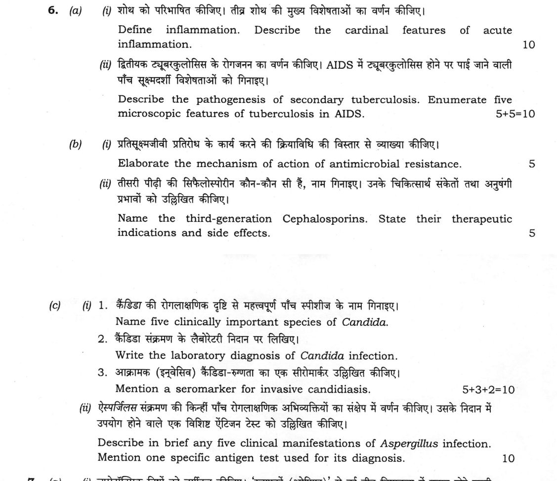
Define inflammation. Describe the cardinal features of acute inflammation. 10 marks
Describe the pathogenesis of secondary tuberculosis. Enumerate five microscopic features of tuberculosis in AIDS. 10 marks
Elaborate the mechanism of action of antimicrobial resistance. 5 marks
Name the third-generation Cephalosporins. State their therapeutic indications and side effects. 5 marks
1. Name five clinically important species of Candida. 2. Write the laboratory diagnosis of Candida infection. 3. Mention a seromarker for invasive candidiasis. 10 marks
Describe in brief any five clinical manifestations of Aspergillus infection. Mention one specific antigen test used for its diagnosis. 10 marks
हिंदी में प्रश्न पढ़ें
शोथ को परिभाषित कीजिए। तीव्र शोथ की मुख्य विशेषताओं का वर्णन कीजिए। (10 अंक)
द्वितीयक ट्यूबरकुलोसिस के रोगजनन का वर्णन कीजिए। AIDS में ट्यूबरकुलोसिस होने पर पाई जाने वाली पाँच सूक्ष्मदर्शी विशेषताओं को गिनाइए। (10 अंक)
प्रतिसूक्ष्मजीवी प्रतिरोध के कार्य करने की क्रियाविधि की विस्तार से व्याख्या कीजिए। (5 अंक)
तीसरी पीढ़ी की सिफैलोस्पोरीन कौन-कौन सी हैं, नाम गिनाइए। उनके चिकित्सार्थ संकेतों तथा अनुपंगी प्रभावों को उल्लिखित कीजिए। (5 अंक)
1. कैंडिडा की रोगलाक्षणिक दृष्टि से महत्वपूर्ण पाँच स्पीशीज़ के नाम गिनाइए। 2. कैंडिडा संक्रमण के लैबोरेटरी निदान पर लिखिए। 3. आक्रामक (इन्वेसिव) कैंडिडा-रुग्णता का एक सीरोमार्कर उल्लिखित कीजिए। (10 अंक)
ऐस्पर्जिलस संक्रमण की किन्हीं पाँच रोगलाक्षणिक अभिव्यक्तियों का संक्षेप में वर्णन कीजिए। उसके निदान में उपयोग होने वाले एक विशिष्ट ऐंटिजन टेस्ट को उल्लिखित कीजिए। (10 अंक)
Model answer
Drafted with AI by UPSC Answer Check against this question's marking rubric, to the expected length. UPSC does not publish answers for Mains — this is one way to score well, not an official key. How we build rubrics and answers.
(a)(i) Inflammation and the cardinal features of acute inflammation (10 marks)
Definition. Inflammation is the local response of living, vascularised tissue to injury (infection, trauma, chemical or immunological damage). It brings plasma proteins and leucocytes to the site to neutralise and remove the injurious agent, and prepares the tissue for repair.
Cardinal features. Celsus described four signs (rubor, calor, tumor, dolor), and Virchow added the fifth (functio laesa). Each is explained by vascular and cellular events that follow injury.
- Rubor (redness): transient arteriolar constriction is followed by vasodilatation and opening of capillary beds under histamine, nitric oxide and prostaglandins. The increased blood flow (active hyperaemia) makes the area red.
- Calor (heat): the same increase in blood flow brings warm blood to the surface, and in a large lesion the pyrogens (IL-1, TNF, PGE₂) add systemic fever.
- Tumor (swelling): increased vascular permeability (endothelial contraction by histamine, bradykinin and leukotrienes, or direct endothelial injury) lets protein-rich fluid escape, the exudate. The high oncotic pressure of the tissue fluid and the cell infiltrate add to the swelling.
- Dolor (pain): bradykinin, prostaglandin E₂ and serotonin stimulate the pain endings, and the pressure of the exudate on the nerve endings adds to it.
- Functio laesa (loss of function): the result of pain, swelling and tissue damage, which limit movement of the part.
Cellular events. Blood flow slows (stasis) as fluid leaves, and leucocytes move to the vessel wall (margination). They roll (selectins), adhere firmly (integrins binding ICAM-1) and cross the wall (diapedesis, using PECAM-1). Under chemotactic signals (C5a, leukotriene B₄, IL-8, bacterial products) they migrate to the site, where they phagocytose the agent and kill it by oxygen-dependent and oxygen-independent mechanisms. Neutrophils dominate for the first 24 hours, and macrophages follow.
(a)(ii) Secondary tuberculosis and TB in AIDS (5 + 5 = 10 marks)
Pathogenesis of secondary (post-primary) tuberculosis. Secondary TB arises in a host already sensitised by primary infection. It follows either the reactivation of dormant bacilli from the primary complex (or another focus), or a fresh exogenous reinfection, usually when immunity is lowered (malnutrition, diabetes, HIV, steroids, alcohol, old age).
- The lesion starts in the apex of the upper lobe (Simon's focus, or the Assmann focus in the infraclavicular region) because the oxygen tension is high there, the organism is strictly aerobic, and the lymph drainage and perfusion are poorer.
- The hypersensitivity (type IV) and acquired immunity shape the lesion. There is a brisk reaction with caseous necrosis, which is contained, so the lesion stays localised, the hilar nodes are spared or only mildly involved, and fibrosis and calcification result.
- Liquefaction of the caseous material and its discharge through a bronchus leaves a cavity, which spreads bacilli by the bronchi (bronchogenic spread) and gives the patient an infectious sputum. Erosion of a vessel causes haemoptysis. Bloodborne spread produces miliary TB, and swallowed sputum causes intestinal disease.
- The lesion may heal by fibrosis and calcification, or progress as fibrocaseous, cavitary disease.
Microscopic features of tuberculosis in AIDS (five). They depend on the CD4 count. In early HIV, the granulomas are typical, but with advanced immunosuppression:
- Poorly formed or absent granulomas, with few epithelioid cells.
- Few Langhans giant cells, and few lymphocytes (loss of CD4 helper cells).
- Extensive necrosis, often not the typical caseation, and sometimes a suppurative (neutrophil-rich) necrosis.
- Numerous acid-fast bacilli in the necrotic areas and inside macrophages on Ziehl–Neelsen stain.
- Sheets of foamy macrophages full of bacilli, with a diffuse infiltrate in place of nodules (and infection with atypical mycobacteria such as M. avium complex in the same pattern).
(b)(i) Mechanisms of antimicrobial resistance (5 marks)
Biochemical mechanisms.
- Enzymatic inactivation of the drug: β-lactamases (penicillin resistance in staphylococci, extended-spectrum β-lactamases and carbapenemases such as NDM-1 and KPC in Gram-negative bacilli), aminoglycoside-modifying enzymes, and chloramphenicol acetyltransferase.
- Altered target site: PBP2a in MRSA (mecA gene), the D-Ala-D-Lac terminus in vancomycin-resistant enterococci, altered DNA gyrase (fluoroquinolones), methylated ribosomal RNA (erm gene, macrolides), and altered RNA polymerase (rifampicin).
- Reduced permeability: loss of outer-membrane porins, as in carbapenem-resistant Pseudomonas.
- Active efflux: pumps expel the drug, as with tetracycline and fluoroquinolones (NorA) and the multidrug MexAB-OprM pump of Pseudomonas.
- Metabolic bypass: altered dihydropteroate synthase or a resistant dihydrofolate reductase (sulphonamides and trimethoprim), and biofilm formation.
Genetic basis. Resistance may be intrinsic, or acquired by chromosomal mutation (for example rifampicin resistance, which is selected by drug pressure), or by horizontal gene transfer. Horizontal transfer is by conjugation (R plasmids, transposons, integrons carrying several genes), transduction (by bacteriophage) and transformation (uptake of naked DNA). Misuse and overuse of antibiotics select the resistant strains.
(b)(ii) Third-generation cephalosporins (5 marks)
Names.
- Parenteral: cefotaxime, ceftriaxone, ceftazidime, cefoperazone and ceftizoxime.
- Oral: cefixime, cefpodoxime proxetil, cefdinir, cefditoren pivoxil and ceftibuten.
Therapeutic indications. Meningitis (ceftriaxone, cefotaxime), gonorrhoea (single dose of ceftriaxone), typhoid (enteric) fever, severe sepsis and Gram-negative bacteraemia, community-acquired pneumonia, complicated urinary tract infection, spontaneous bacterial peritonitis, Lyme disease and surgical prophylaxis. Ceftazidime and cefoperazone are used against Pseudomonas aeruginosa, including in neutropenic fever.
Side effects. Hypersensitivity (cross-reaction with penicillin in about 5–10%), diarrhoea and superinfection (Clostridioides difficile, candidiasis), thrombophlebitis, biliary sludge and pseudolithiasis (ceftriaxone), hypoprothrombinaemia with bleeding and a disulfiram-like reaction with alcohol (cefoperazone), neutropenia and haemolytic anaemia, and raised liver enzymes. Ceftriaxone is contraindicated in neonates who need calcium-containing infusions (precipitation) and in hyperbilirubinaemic neonates.
(c)(i) Candida (5 + 3 + 2 = 10 marks)
1. Five clinically important species (5 marks). Candida albicans (the commonest), C. tropicalis, C. glabrata, C. parapsilosis and C. krusei. C. auris is an emerging, multidrug-resistant species, and C. dubliniensis is closely related to C. albicans.
2. Laboratory diagnosis (3 marks).
- Specimens: swabs and scrapings, sputum, urine, blood, CSF, tissue and catheter tips.
- Microscopy: KOH mount or Gram stain shows oval budding yeast cells (3–6 µm) with pseudohyphae. Tissue is stained with PAS or silver.
- Culture: Sabouraud dextrose agar at 25–37 °C gives creamy white, pasty colonies. Chromogenic agar (CHROMagar) separates species by colour (C. albicans green). Blood culture is used for candidaemia.
- Identification: the germ tube test (positive for C. albicans in serum at 37 °C in 2–3 hours), chlamydospore formation on corn meal agar, sugar assimilation and fermentation tests, MALDI-TOF and PCR.
3. Seromarker of invasive candidiasis (2 marks). Serum (1→3)-β-D-glucan, a cell wall component, is the marker (with mannan antigen and anti-mannan antibody as an alternative). It is not specific for Candida.
(c)(ii) Aspergillus infection (10 marks)
Clinical manifestations (any five).
- Allergic bronchopulmonary aspergillosis (ABPA): a type I and type III hypersensitivity in asthmatics and cystic fibrosis, with wheeze, eosinophilia, raised IgE and fleeting infiltrates.
- Aspergilloma (fungal ball): saprophytic growth in a pre-existing lung cavity (old tuberculosis), causing haemoptysis.
- Invasive pulmonary aspergillosis: in neutropenic and transplant patients, with fever, pleuritic pain, haemoptysis and angioinvasion (halo sign on CT), and dissemination to the brain.
- Chronic pulmonary aspergillosis: chronic cavitary and necrotising disease in a lung with structural damage.
- Extrapulmonary: allergic or invasive fungal sinusitis, otomycosis, fungal keratitis, cutaneous infection and endocarditis. Aspergillus flavus also causes aflatoxin poisoning and liver cancer.
Specific antigen test. The galactomannan assay, an ELISA for the cell wall polysaccharide galactomannan in serum or bronchoalveolar lavage fluid (positive at an index of 0.5 or more in serum), is used for invasive aspergillosis. False positives occur with some antibiotics such as piperacillin–tazobactam.
How this answer will be evaluated
Evaluation rubric
Each sub-part is marked on its own, against the marks and word limit printed on the paper.
- (a)(i) (a)(i) Define inflammation and describe the cardinal features of acute inflammation (10 marks). 10 marks
Must cover
- Definition: local response of vascularised tissue to injury, bringing cells and plasma proteins
- Rubor and calor from vasodilatation and increased blood flow (histamine, NO, prostaglandins)
- Tumor from increased vascular permeability and exudate; dolor from bradykinin and prostaglandins; functio laesa
- Cellular events: margination, rolling, adhesion, diapedesis, chemotaxis and phagocytosis
Loses marks
- Listing the signs without mechanisms.
- Describing chronic inflammation instead.
- Omitting functio laesa.
Earns more
- Mechanism explained for each sign, not just named.
- Latin names of all five signs, with Virchow's addition.
Extra mark
- Systemic effects (fever, leucocytosis) and mediators
- (a)(ii) (a)(ii) Describe the pathogenesis of secondary tuberculosis and enumerate five microscopic features of TB in AIDS (5 + 5 = 10 marks). 10 marks
Must cover
- Reactivation or reinfection in a sensitised host, precipitated by immunosuppression
- Apical location from high oxygen tension and poor lymph drainage
- Type IV hypersensitivity: caseation, cavitation and bronchogenic spread with hilar nodes spared
- AIDS: poorly formed or absent granulomas, few giant cells and lymphocytes, extensive necrosis and numerous acid-fast bacilli
Loses marks
- Describing primary tuberculosis.
- Stating that granulomas are well formed in advanced AIDS.
- Fewer than five microscopic features.
Earns more
- Explanation why the apex is involved.
- Five features stated for TB in AIDS.
Extra mark
- Simon's and Assmann foci; atypical mycobacteria
- (b)(i) (b)(i) Elaborate the mechanism of action of antimicrobial resistance (5 marks). 5 marks
Must cover
- Enzymatic inactivation (β-lactamase, aminoglycoside-modifying enzymes)
- Altered target (PBP2a in MRSA, D-Ala-D-Lac in VRE, gyrase, ribosomal methylation)
- Reduced permeability (porin loss) and active efflux
- Genetic basis: mutation and horizontal transfer by conjugation, transduction, transformation
Loses marks
- Describing how the drug acts instead of how resistance arises.
- Omitting the genetic basis.
- Giving only one mechanism.
Earns more
- A named drug–organism example for each mechanism.
- Plasmids, transposons and integrons mentioned.
Extra mark
- Metabolic bypass and biofilm formation
- (b)(ii) (b)(ii) Name the third-generation cephalosporins and state their therapeutic indications and side effects (5 marks). 5 marks
Must cover
- Parenteral: cefotaxime, ceftriaxone, ceftazidime, cefoperazone
- Oral: cefixime, cefpodoxime, cefdinir
- Indications: meningitis, gonorrhoea, typhoid fever, sepsis, Pseudomonas (ceftazidime), pneumonia
- Side effects: hypersensitivity, diarrhoea and superinfection, biliary sludge (ceftriaxone), bleeding and disulfiram-like reaction (cefoperazone)
Loses marks
- Naming first- or second-generation drugs as third generation.
- Omitting side effects.
- Stating that they have no activity against Gram-negative bacilli.
Earns more
- Several drugs named in both groups.
- Drug-specific effects stated.
Extra mark
- Neonatal contraindications of ceftriaxone
- (c)(i) (c)(i) Name five clinically important Candida species; write the laboratory diagnosis of Candida infection; mention a seromarker for invasive candidiasis (5 + 3 + 2 = 10 marks). 10 marks
Must cover
- Species: C. albicans, C. tropicalis, C. glabrata, C. parapsilosis, C. krusei (or C. auris)
- Specimens and direct microscopy showing budding yeast with pseudohyphae
- Culture on Sabouraud agar and CHROMagar; germ tube test, chlamydospores, sugar tests, MALDI-TOF
- Seromarker: (1→3)-β-D-glucan (or mannan antigen)
Loses marks
- Naming Aspergillus or Cryptococcus species.
- Giving galactomannan as the Candida marker.
- Omitting the culture medium.
Earns more
- Five species named correctly.
- Germ tube test details.
Extra mark
- C. auris as an emerging multidrug-resistant species
- (c)(ii) (c)(ii) Describe in brief any five clinical manifestations of Aspergillus infection; mention one specific antigen test used for diagnosis (10 marks). 10 marks
Must cover
- Five manifestations from ABPA, aspergilloma, invasive pulmonary aspergillosis, chronic pulmonary aspergillosis, sinusitis, otomycosis, keratitis
- ABPA with asthma, eosinophilia and raised IgE
- Aspergilloma in a pre-existing cavity with haemoptysis; invasive disease in neutropenia with the halo sign
- Galactomannan antigen test (ELISA on serum or BAL)
Loses marks
- Giving β-D-glucan as the specific antigen.
- Fewer than five manifestations.
- Describing only laboratory findings.
Earns more
- Brief description of each manifestation with its setting.
- Antigen named with the sample and index cut-off.
Extra mark
- Aflatoxin poisoning by A. flavus; false positives with some antibiotics
What "Describe" is asking you to do
Give a full, ordered account of the thing named — its parts, stages or mechanism — in the sequence in which it actually exists or occurs. Most describe questions come from the science optionals, where the marks sit in correct technical detail and, where the stem says so, a labelled diagram.
Structure that answers it
One-line identification of the subject → the parts or stages in their real order, each with its defining detail → labelled diagram where the subject is structural → closing line on function or significance
Where marks are lost
Loose general prose where the examiner is ticking named parts, correct terminology and their sequence; and in the General Studies papers, turning to evaluation before the description is finished.
Practice this exact question
Write your answer and it is marked point by point against the model answer above — what you covered, what you missed, what you got wrong.
Evaluate my answer →More from Medical Science 2026 Paper I
- Q3 (a) (i) 1. Name the ligaments of ovary and their role in ovarian torsion and its complica…
- Q4 (a) (i) Describe the cerebrospinal fluid (CSF) flow from site of production to its absorp…
- Q5 (a) (i) State the therapeutic indications, drug interactions and side effects of Methotre…
- Q6 (a) (i) Define inflammation. Describe the cardinal features of acute inflammation. (10 ma…
- Q7 (a) (i) Classify neurotoxic poisons. Discuss in brief the clinical features, treatment an…
- Q8 (a) (i) 1. Enumerate five agents of malaria in humans. 2. Describe in brief rapid diagnos…