Paper II — Q3
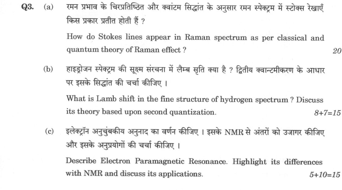
(a) How do Stokes lines appear in Raman spectrum as per classical and quantum theory of Raman effect ? 20 marks (b) What is Lamb…
How do Stokes lines appear in Raman spectrum as per classical and quantum theory of Raman effect ? 20 marks
What is Lamb shift in the fine structure of hydrogen spectrum ? Discuss its theory based upon second quantization. 8+7=15 marks
Describe Electron Paramagnetic Resonance. Highlight its differences with NMR and discuss its applications. 5+10=15 marks
हिंदी में प्रश्न पढ़ें
रमन प्रभाव के चिरप्रतिष्ठित और क्वांटम सिद्धांत के अनुसार रमन स्पेक्ट्रम में स्टोक्स रेखाएं किस प्रकार प्रतीत होती हैं ? 20 अंक
हाइड्रोजन स्पेक्ट्रम की सूक्ष्म संरचना में लैम्ब सृति क्या है ? द्वितीय क्वांटमीकरण के आधार पर इसके सिद्धांत की चर्चा कीजिए । 8+7=15 अंक
इलेक्ट्रॉन अनुचुंबकीय अनुनाद का वर्णन कीजिए । इसके NMR से अंतरों को उजागर कीजिए और इसके अनुप्रयोगों की चर्चा कीजिए । 5+10=15 अंक
Model answer
Written by UPSC Answer Check against this question's marking rubric, to the expected length. UPSC does not publish answers for Mains — this is one way to score well, not an official key.
Raman effect. In the classical picture, an incident field E=E0 cos ωt makes the molecular polarizability oscillate as α=α0+(∂α/∂Q)Q, with Q=Q0 cos ω_v t. The induced dipole p=αE contains frequencies ω, ω±ω_v. The ω term gives Rayleigh scattering; ω−ω_v gives Stokes lines, lower in frequency because the molecule gains vibrational energy; ω+ω_v gives anti-Stokes lines. The scattered intensity is proportional to (∂α/∂Q)², so only vibrations that modulate polarizability are seen. The classical result explains the frequency shifts but not the detailed intensity ratio; quantum theory supplies the populations and selection rules. C.V. Raman’s 1928 Calcutta experiment used a mercury arc, polarizers, a liquid sample and a spectrograph to observe these weak shifted lines.
Quantum mechanically, scattering is a second-order process through virtual intermediate states, not real absorption. The virtual states are off-resonant, so energy conservation is satisfied by the molecular vibrational change. The Kramers–Heisenberg dispersion formula gives the amplitude as a sum over states n of products of dipole matrix elements divided by energy denominators, with both emission-then-absorption and absorption-then-emission terms. Placzek’s polarizability theory reduces the intensity to the square of the relevant polarizability tensor component, α_ij (α_xx, α_xy, α_zz, etc.), so a vibration is Raman active only if α_ij changes with the normal coordinate; for a harmonic oscillator Δv=±1. In centrosymmetric molecules, modes that change dipole moment are IR active while modes that change polarizability are Raman active, giving mutual exclusion. The Stokes/anti-Stokes ratio is I_S/I_AS ≈ (ν_S/ν_AS)^4 (N_v+1)/N_v ≈ (ν_S/ν_AS)^4 exp(hν_v/kT), where N_v is the mean vibrational occupation; hence Stokes lines dominate at ordinary temperatures.
Lamb shift. Dirac theory predicts degeneracy of hydrogen 2S1/2 and 2P1/2, but Lamb and Retherford (1947) found 2S1/2 higher by about 1058 MHz. Bethe’s calculation treated the electron’s interaction with the quantized radiation field and introduced mass renormalization to remove the self-energy divergence. In second quantization, the Dirac and electromagnetic fields are expanded in creation and annihilation operators, and the interaction Hamiltonian is H_int=−e∫ψbar γ^μ A_μ ψ d³x. This allows the electron to emit and reabsorb a virtual photon, represented by the one-loop self-energy Feynman diagram; vacuum fluctuations and radiation reaction shift the energy levels. The one-loop correction is integrated over virtual photon momentum; its divergence is absorbed into the electron mass, while the finite remainder gives the shift. The bare mass and charge are divergent, so renormalization fixes them to observed values, leaving a finite Lamb shift that confirms QED.
EPR. Electron Paramagnetic Resonance detects transitions between electron spin sublevels in a magnetic field. The electron spin magnetic moment is μ=−g μ_B S/h; in B0 the m_s=±1/2 levels split. For an unpaired electron, H=μ_B g S·B/h, giving ΔE=gμ_BB and resonance hν=gμ_BB, or B0=hν/(gμ_B). Hyperfine coupling, H_hf=A I·S, splits the line and identifies nuclear spin and coupling constants; in solids the g-factor is anisotropic, giving orientation-dependent resonance. In powders, anisotropy produces broadened spectra; in single crystals, orientation can be resolved. EPR uses microwave frequencies and lower fields because the electron magnetic moment is much larger than the nuclear magneton.
| Feature | EPR | NMR | |---|---|---| | Magnetic moment | μ_B, ~658 μ_N | μ_N | | Typical field | ~0.3 T (X-band) | ~10 T | | Relaxation | shorter T1/T2, spin–orbit/dipolar | longer, weaker coupling | | Sample | paramagnetic centres, small/frozen | abundant nuclei, larger/high field | | Sensitivity | very sensitive to unpaired e⁻ | bulk nuclei, needs high concentration |
Applications include ESR dating of archaeological samples such as Bhimbetka rock paintings, where trapped unpaired electrons record accumulated radiation dose; detection of radical pairs in photosynthetic reaction centres; Gd³⁺ MRI contrast agents that shorten water-proton T1; and spin labels for protein structure through dipolar distance measurements. These applications show EPR’s value in archaeology, biochemistry, medicine and structural biology.
Thus Raman scattering, Lamb shift and EPR respectively reveal molecular polarizability, QED self-energy corrections and electron spin resonance; each connects a microscopic interaction to a measurable spectral shift.
What "Explain" is asking you to do
Make the working of something clear — what sets it off, what follows from what, and what it produces. Explain is the Commission's mechanism word: it dominates the technical papers and the “explain why” stems, where the marks sit in the causal chain and not in the label.
Structure that answers it
State what it is → the initiating condition → the chain of cause, step by step → an instance where it plays out → what the chain produces
Where marks are lost
Describing what something looks like instead of why it works that way. Naming the stages without linking them reads as description too.
How this answer will be evaluated
Approach
Framework: Principle > Setup and diagram > Derivation > Result and limiting case. (a) explain: definition/context > points in order > small example > short close | (b) discuss: intro > 3-4 dimensions > example > balanced close | (c) describe: define > structure or process in order > labelled diagram > significance Full marks: Rigorous derivation, clear diagrams, and physical insight.
Key points expected
- Classical theory: induced dipole moment and beat frequency
- Quantum theory: energy level diagram with virtual state
- Derivation of frequency shift for Stokes line
- Physical interpretation of energy loss to molecule
- Definition of Lamb shift in fine structure
- Second quantization formalism for interaction
- Self-energy and vacuum polarization contributions
- Calculation of energy shift for 2S1/2 and 2P1/2
Evaluation rubric
Each sub-part is marked on its own, against the marks and word limit printed on the paper.
- (a) Explain appearance of Stokes lines via classical and quantum theories. 20 marks
explain— definition/context → points in order → small example → short close
Must cover
- Classical theory: induced dipole moment and beat frequency
- Quantum theory: energy level diagram with virtual state
- Derivation of frequency shift for Stokes line
- Physical interpretation of energy loss to molecule
Loses marks
- Formula substitution without derivation
- Confusing Raman with Compton scattering
Earns more
- Labelled energy level diagram
- Mention of anti-Stokes lines for contrast
- Selection rules for Raman activity
Extra mark
- Comparison of intensity of Stokes vs anti-Stokes
- (b) Define Lamb shift and discuss theory based on second quantization. 15 marks
discuss— intro → 3-4 dimensions → example → balanced close
Must cover
- Definition of Lamb shift in fine structure
- Second quantization formalism for interaction
- Self-energy and vacuum polarization contributions
- Calculation of energy shift for 2S1/2 and 2P1/2
Loses marks
- Treating as a classical perturbation
- Ignoring the role of virtual photons
Earns more
- Mention of Bethe's non-relativistic calculation
- Role of renormalization in QED
Extra mark
- Numerical value of the shift (1057 MHz)
- (c) Describe EPR, highlight differences with NMR, and discuss applications. 15 marks
describe— define → structure or process in order → labelled diagram → significance
Must cover
- Principle of EPR: electron spin resonance
- Resonance condition: hν = gμB B
- Differences with NMR: frequency, g-factor, sensitivity
- Applications: free radicals, transition metal complexes
Loses marks
- Confusing electron and nuclear magnetic moments
- Listing applications without context
Earns more
- Labelled energy level diagram for electron spin
- Mention of hyperfine structure in EPR
Extra mark
- Specific example of a radical studied by EPR
Practice this exact question
Write your answer and it is marked point by point against the model answer above — what you covered, what you missed, what you got wrong.
Evaluate my answer →More from Physics 2024 Paper II
- Q1 Q1. (a) A particle limited to the x-axis has the wave function φ(x) = bx² between x = 0 a…
- Q2 Q2. (a) Prove that : (i) [L², Lz] = 0 (ii) [Lz, L+] = ℏL+ (iii) [L+, L-] = 2ℏLz (iv) L+ L…
- Q3 (a) How do Stokes lines appear in Raman spectrum as per classical and quantum theory of R…
- Q4 (a) (i) Using free electron theory of metals, calculate the Fermi energy level of sodium…
- Q5 (a) Compare nuclear density of hydrogen (₁H¹) with its atomic density. (Assume the atom t…
- Q6 (a) Does the nucleus possess magnetic moment ? Justify your answer. Define nuclear magnet…