Paper II — Q6
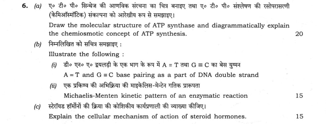
(a) Draw the molecular structure of ATP synthase and diagrammatically explain the chemiosmotic concept of ATP synthesis. 20 (b)…
Draw the molecular structure of ATP synthase and diagrammatically explain the chemiosmotic concept of ATP synthesis. 20 marks
Illustrate the following :
A = T and G ≡ C base pairing as a part of DNA double strand
Michaelis-Menten kinetic pattern of an enzymatic reaction 15
Explain the cellular mechanism of action of steroid hormones. 15 marks
हिंदी में प्रश्न पढ़ें
ए० टी० पी० सिन्थेज की आण्विक संरचना का चित्र बनाइए तथा ए० टी० पी० संश्लेषण की रसोप्रसरणी (केमिऑस्मोटिक) संकल्पना को आरेखीय रूप से समझाइए। 20
निम्नलिखित को सचित्र समझाइए :
डी० एन० ए० द्वैलड़ी के एक भाग के रूप में A = T तथा G ≡ C का बेस युग्मन
एक प्रकिण्व की अभिक्रिया की माइकेलिस-मेंटेन गतिक प्राक्रता 15
स्टेरॉयड हार्मोनों की क्रिया की कोशिकीय कार्यप्रणाली की व्याख्या कीजिए। 15
Model answer
Written by UPSC Answer Check against this question's marking rubric, to the expected length. UPSC does not publish answers for Mains — this is one way to score well, not an official key.
(a) ATP synthase is a rotary molecular motor of the inner mitochondrial membrane, chloroplast thylakoid and bacterial plasma membrane. Schematic molecular structure:
Intermembrane side (high H⁺): H⁺ → a-subunit half-channels Membrane-embedded F₀: a, b₂, c-ring (c₈–c₁₂ in different species) Stalk: central γε rotor; peripheral stalk b₂δ/OSCP Matrix side F₁: α₃β₃ hexamer with catalytic β sites Matrix: ADP + Pi → ATP
F₀ conducts protons; F₁ is catalytic. The γε stalk connects them. The β subunits cycle through O (open), L (loose) and T (tight) conformations.
Chemiosmotic concept: Peter Mitchell proposed that electron transport chains I, III and IV pump H⁺ from the matrix to the intermembrane space. This creates a proton motive force. In standard form, Δp = Δψ − (2.303RT/F)ΔpH, where Δψ = ψ_out − ψ_in and ΔpH = pH_out − pH_in. In mitochondria, ψ_out > ψ_in, so Δψ is positive; H⁺ is pumped out, so pH_out < pH_in, making ΔpH negative, and the chemical term also favours H⁺ entry. R is in J mol⁻¹ K⁻¹, T is in K and F is in C mol⁻¹; Δψ and RT/F must be in the same units. Protons re-enter through F₀ down the gradient. Protonation/deprotonation of c-ring carboxylates and a-subunit half-channels rotate the c-ring. The γε rotor rotates inside α₃β₃, changing each β subunit O→L→T→O. In L, ADP and Pi bind; in T, ADP + Pi condense to ATP; in O, ATP is released. Typically about 3–4 H⁺ are used per ATP synthesised, depending on c-ring stoichiometry, plus about 1 H⁺ for ADP/ATP antiport. Validity: intact proton-impermeable inner membrane, functional ETC, O₂ as terminal acceptor, ADP + Pi, and Mg²⁺.
(b) Illustrate the following:
(i) In B-DNA, two antiparallel strands pair by Watson–Crick hydrogen bonds: A=T has two H bonds, and G≡C has three H bonds. Purine pairs with pyrimidine, keeping helix width ~2 nm.
5′—A—G—C—T—3′ = ≡ ≡ = 3′—T—C—G—A—5′
Thus A on one strand pairs only with T on the other; G only with C. G≡C has one extra H bond, increasing thermal stability. Bases stack hydrophobically; the phosphodiester backbone lies outside. Condition: right-handed B-DNA under normal ionic strength and pH.
(ii) Michaelis–Menten kinetics describes a single-substrate enzyme: E + S ⇌ ES → E + P. Let k₁, k₋₁ and k₂ be rate constants. At steady state, d[ES]/dt = 0: k₁[E][S] = (k₋₁ + k₂)[ES]. With [E]total = [E] + [ES], solving gives: [ES] = [E]total[S]/(Kₘ + [S]), where Kₘ = (k₋₁ + k₂)/k₁. Initial rate v₀ = k₂[ES], so: v₀ = Vₘₐₓ[S]/(Kₘ + [S]), with Vₘₐₓ = k₂[E]total.
Graph: v₀ versus [S] is a rectangular hyperbola. v₀ | __________ Vₘₐₓ | / | / | / |__/________________ [S] Kₘ at Vₘₐₓ/2
At low [S] (S << Kₘ), v₀ ≈ (Vₘₐₓ/Kₘ)[S], first-order. At high [S] (S >> Kₘ), v₀ ≈ Vₘₐₓ, zero-order. Kₘ is the substrate concentration at v₀ = Vₘₐₓ/2; unit mol L⁻¹. Vₘₐₓ has unit mol L⁻¹ s⁻¹ or µmol min⁻¹ mg⁻¹. Linear form: 1/v₀ = (Kₘ/Vₘₐₓ)(1/[S]) + 1/Vₘₐₓ. Conditions: [S] >> [E], initial velocity, steady state, no cooperativity, and negligible product.
(c) Steroid hormones are lipophilic cholesterol derivatives such as cortisol, aldosterone, testosterone, estradiol and progesterone. They cross the plasma membrane by simple diffusion. In target cells they bind intracellular receptors—cytoplasmic or nuclear. Unbound receptor is complexed with heat-shock proteins HSP90, HSP70, p23 and immunophilins, which mask the DNA-binding domain and nuclear localization signals.
Hormone binding causes a conformational change, dissociation of HSPs, receptor dimerisation and exposure of a zinc-finger DNA-binding domain. The hormone–receptor complex enters the nucleus and binds to hormone response elements (HREs) in promoter or enhancer regions of target genes. It recruits coactivators or corepressors and alters RNA polymerase II activity. This changes transcription, mRNA processing and protein synthesis. Effects are delayed (hours) but prolonged. Example: cortisol–glucocorticoid receptor activates PEPCK; aldosterone–mineralocorticoid receptor induces Na⁺/K⁺-ATPase; estrogen–estrogen receptor promotes cell proliferation. Some steroids also act rapidly at membrane receptors via second messengers. Termination occurs by metabolic inactivation and excretion. Validity: requires functional receptor, HRE, chromatin accessibility and cofactors.
What "Draw" is asking you to do
Produce the diagram as the answer, not as an ornament to it. Where the question lists several items — circuit, function table, logic symbol, structure — each is separately marked, and the lines of text must refer to the diagram through its own labels.
Structure that answers it
Diagram drawn large and clean → every part, axis and terminal labelled → caption → two or three lines tying it to what was asked
Where marks are lost
Delivering part of the list and leaving the rest, which forfeits those marks directly. In chemistry, a flat sketch where the geometry or stereochemistry was the point; in engineering, unlabelled terminals, missing polarity, or no sign convention stated.
How this answer will be evaluated
Approach
Framework: Zoology, Paper 2. (a) explain: definition/context > points in order > small example > short close | (b(i)) describe: define > structure or process in order > labelled diagram > significance | (b(ii)) describe: define > structure or process in order > labelled diagram > significance | (c) explain: definition/context > points in order > small example > short close Full marks: Precise diagrams with all labels; clear mechanistic steps; correct terminology.
Key points expected
- Labelled diagram of F1 and Fo subunits
- Explanation of proton gradient (H+) driving rotation
- Mechanism of ADP + Pi to ATP conversion
- Link to electron transport chain (ETC)
- Diagram showing antiparallel strands
- Correct hydrogen bonding (2 for A-T, 3 for G-C)
- Labeling of nitrogenous bases
- Indication of sugar-phosphate backbone
Evaluation rubric
Each sub-part is marked on its own, against the marks and word limit printed on the paper.
- (a) Molecular structure of ATP synthase and chemiosmotic mechanism of ATP synthesis. 20 marks
explain— definition/context → points in order → small example → short close
Must cover
- Labelled diagram of F1 and Fo subunits
- Explanation of proton gradient (H+) driving rotation
- Mechanism of ADP + Pi to ATP conversion
- Link to electron transport chain (ETC)
Loses marks
- Diagram without labels
- Confusing ATP synthase with ATPase
- Omitting the role of proton motive force
Earns more
- Mention of Mitchell's chemiosmotic hypothesis
- Specific subunit names (alpha, beta, gamma, c-ring)
- Diagram showing rotation of gamma subunit
Extra mark
- Mention of specific stoichiometry (H+ per ATP)
- (b(i)) Diagrammatic illustration of A=T and G≡C base pairing in DNA.
describe— define → structure or process in order → labelled diagram → significance
Must cover
- Diagram showing antiparallel strands
- Correct hydrogen bonding (2 for A-T, 3 for G-C)
- Labeling of nitrogenous bases
- Indication of sugar-phosphate backbone
Loses marks
- Incorrect number of hydrogen bonds
- Parallel strands instead of antiparallel
Earns more
- Mention of Chargaff's rules
- Clear distinction between purines and pyrimidines
Extra mark
- Mention of major/minor grooves
- (b(ii)) Michaelis-Menten kinetic pattern of an enzymatic reaction.
describe— define → structure or process in order → labelled diagram → significance
Must cover
- Graph of Velocity (V) vs Substrate ([S])
- Identification of Vmax and Km
- Explanation of saturation kinetics
- Equation: V = Vmax[S] / (Km + [S])
Loses marks
- Graph without axes labels
- Confusing Km with Vmax
Earns more
- Mention of Lineweaver-Burk plot
- Explanation of Km as affinity constant
Extra mark
- Mention of kcat (turnover number)
- (c) Cellular mechanism of action of steroid hormones. 15 marks
explain— definition/context → points in order → small example → short close
Must cover
- Lipophilic nature allowing membrane diffusion
- Intracellular receptor binding (cytoplasm/nucleus)
- Hormone-receptor complex acting as transcription factor
- Gene transcription and protein synthesis
Loses marks
- Describing cell surface receptor mechanism
- Omitting the genomic effect (transcription)
Earns more
- Comparison with peptide hormones (second messengers)
- Mention of specific steroid (e.g., cortisol, testosterone)
Extra mark
- Mention of specific gene targets
Practice this exact question
Write your answer and it is marked point by point against the model answer above — what you covered, what you missed, what you got wrong.
Evaluate my answer →More from Zoology 2022 Paper II
- Q3 (a) Sketch out a basic transcription unit and illustrate the RNA polymerase II associated…
- Q4 (a) What is fossil? Describe the origin and evolution of horse based on the fossil record…
- Q5 Write your answer in about 150 words for each of the following : 10×5=50 (a) Define immun…
- Q6 (a) Draw the molecular structure of ATP synthase and diagrammatically explain the chemios…
- Q7 (a) What are intestinal glands? Describe the roles of intestinal juices in digestion and…
- Q8 (a) What is stem cell? Discuss the types of stem cells and their application in therapeut…