Paper II — Q2
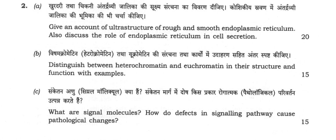
(a) Give an account of ultrastructure of rough and smooth endoplasmic reticulum. Also discuss the role of endoplasmic reticulum…
Give an account of ultrastructure of rough and smooth endoplasmic reticulum. Also discuss the role of endoplasmic reticulum in cell secretion. 20 marks
Distinguish between heterochromatin and euchromatin in their structure and function with examples. 15 marks
What are signal molecules? How do defects in signalling pathway cause pathological changes? 15 marks
हिंदी में प्रश्न पढ़ें
खुरदरी तथा चिकनी अंतर्द्रव्यी जालिका की सूक्ष्म संरचना का विवरण दीजिए। कोशिकीय स्रवण में अंतर्द्रव्यी जालिका की भूमिका की भी चर्चा कीजिए। (20 अंक)
विषमक्रोमैटिन (हेट्रोक्रोमैटिन) तथा युक्रोमैटिन की संरचना तथा कार्यों में उदाहरण सहित अंतर स्पष्ट कीजिए। (15 अंक)
संकेत अणु (सिग्नल मॉलिक्यूल) क्या हैं? संकेत मार्ग में दोष किस प्रकार रोगात्मक (पैथोलॉजिकल) परिवर्तन उत्पन्न करते हैं? (15 अंक)
Model answer
Written by UPSC Answer Check against this question's marking rubric, to the expected length. UPSC does not publish answers for Mains — this is one way to score well, not an official key.
The eukaryotic endomembrane system, nuclear architecture, and intercellular communication networks operate in close coordination to regulate cellular homeostasis and physiological function.
Ultrastructure and Secretory Role of Endoplasmic Reticulum
The endoplasmic reticulum (ER) exists as two morphologically and functionally distinct domains: the rough endoplasmic reticulum (RER) and the smooth endoplasmic reticulum (SER). Ultrastructurally, the RER comprises an extensive network of parallel, flattened, membrane-delimited saccules termed cisternae. The cytosolic surface of the RER membrane is studded with 80S ribosomes bound via translocon complexes and ribophorins I and II, enclosing a continuous luminal space. In contrast, the SER consists of a convoluted, three-dimensional meshwork of interconnected tubules devoid of membrane-bound ribosomes, maintaining continuity with the RER.
The ER plays a pivotal role in cellular secretion through an integrated biosynthetic pathway. Secretory proteins bearing an N-terminal hydrophobic signal peptide are recognized by the Signal Recognition Particle (SRP) and translocated cotranslationally into the RER lumen. Within the lumen, molecular chaperones like BiP (Binding immunoglobulin Protein) and calnexin ensure proper protein folding and disulfide bond formation, while oligosaccharyltransferase initiates core N-linked glycosylation. Misfolded proteins are exported and degraded via ER-associated degradation (ERAD). Correctly folded secretory proteins are packaged into COPII-coated transport vesicles that bud from ER exit sites and fuse with the cis-Golgi network for post-translational modification, sorting, and eventual exocytic release. Concurrently, the SER facilitates secretion by synthesizing membrane lipids, lipoproteins, and steroid hormones, while also housing cytochrome P450 monooxygenases for metabolic detoxification.
Structural and Functional Distinction of Chromatin Domains
Nuclear chromatin is compartmentalized into euchromatin and heterochromatin, reflecting dynamic states of gene expression:
Structurally, euchromatin exists in a relaxed, decondensed 10 to 30 nm nucleosomal fiber conformation during interphase. It replicates early in the S-phase and is characterized by hyperacetylated histones and hypomethylated DNA. Functionally, euchromatin is transcriptionally active, allowing access to RNA polymerase II and transcription factors. Examples include ubiquitously expressed housekeeping genes, such as GAPDH and actin, as well as developmentally inducible genes.
Heterochromatin remains highly condensed and cytologically dense throughout interphase. It replicates late in the S-phase and exhibits epigenetic marks such as histone H3 lysine 9 trimethylation (H3K9me3) and hypermethylated DNA, rendering it transcriptionally silent. It is categorized into two forms: Constitutive heterochromatin remains permanently silenced across all cell types, serving structural roles at centromeres and telomeres to maintain chromosomal integrity. Facultative heterochromatin possesses the capacity to alternate between condensed and relaxed states depending on developmental signals. A classic mammalian example is dosage compensation via X-chromosome inactivation (lyonization), where one female X-chromosome condenses into a transcriptionally inert Barr body.
Signal Molecules and Pathological Manifestations of Pathway Defects
Signal molecules, or ligands, are chemical entities that mediate intercellular communication via endocrine, paracrine, autocrine, or juxtacrine modes. They include peptide hormones (insulin, glucagon), steroid hormones (estrogen, cortisol), growth factors (EGF, FGF), cytokines (interleukins), and neurotransmitters (acetylcholine, dopamine).
Defects occurring at distinct nodes of signaling cascades disrupt cellular homeostasis, generating severe pathological conditions: First, receptor mutations and overexpression lead to aberrant downstream firing. Gain-of-function mutations in the Epidermal Growth Factor Receptor (EGFR) drive continuous cell survival and proliferation in non-small cell lung carcinomas and glioblastomas. Second, dysregulation of heterotrimeric G-proteins disrupts intracellular second messengers. For example, the Vibrio cholerae enterotoxin covalently modifies the Gαs subunit via ADP-ribosylation, locking it in an active state; this causes sustained adenylate cyclase activation, hyper-elevated cAMP, and severe secretory diarrhea. Conversely, Bordetella pertussis toxin inactivates Gαi, impairing immune clearance. Third, intracellular kinase cascades are frequently mutated in malignancies. Constitutive activation of the Ras-Raf-MEK-MAPK pathway due to K-Ras mutations bypasses normal growth checkpoints. In India, this pathway is frequently implicated in the high incidence of oral squamous cell carcinoma (OSCC), where betel quid and tobacco carcinogens induce mutations in EGFR, Ras, and cyclin D1 networks.
A comprehensive mapping of ER proteostasis, chromatin dynamics, and signal transduction cascades is essential for advancing targeted molecular therapies, including kinase inhibitors and epigenetic drugs, to correct these pathological perturbations.
What "Discuss" is asking you to do
Lay the issue out from more than one side — how it arose, what is claimed for it, what is held against it, and where it now stands. UPSC attaches discuss to broad topics with several live dimensions, so coverage of the dimensions earns more than the strength of your opinion.
Structure that answers it
Set the issue up → the case as it is made → the case against → the dimension both sides leave out → where the balance now lies
Where marks are lost
Listing facts with no thread between them, or arguing one side throughout and calling it a discussion.
How this answer will be evaluated
Approach
(a) discuss: intro > 3-4 dimensions > example > balanced close | (b) compare: paired headings or table > key differences > significance > conclusion | (c) explain: definition/context > points in order > small example > short close Full marks: Comprehensive, accurate, with specific examples and clear mechanisms.
Key points expected
- RER: ribosomes, cisternae, rough appearance
- SER: smooth, tubular, no ribosomes
- Secretion: protein synthesis, folding, transport
- Link to Golgi apparatus and vesicles
- Heterochromatin: condensed, transcriptionally inactive
- Euchromatin: decondensed, transcriptionally active
- Structural differences (staining, packing)
- Functional differences (gene expression)
Evaluation rubric
Each sub-part is marked on its own, against the marks and word limit printed on the paper.
- (a) Detailed ultrastructure of RER/SER and their role in secretion. 20 marks
discuss— intro → 3-4 dimensions → example → balanced close
Must cover
- RER: ribosomes, cisternae, rough appearance
- SER: smooth, tubular, no ribosomes
- Secretion: protein synthesis, folding, transport
- Link to Golgi apparatus and vesicles
Loses marks
- Confusing RER and SER functions
- Omitting the role in protein folding
Earns more
- Mention of specific enzymes in SER
- Diagram of ER-Golgi pathway
- Distinction between secretory and membrane proteins
Extra mark
- Mention of specific protein examples (e.g., insulin)
- (b) Distinguish heterochromatin and euchromatin in structure and function. 15 marks
compare— paired headings or table → key differences → significance → conclusion
Must cover
- Heterochromatin: condensed, transcriptionally inactive
- Euchromatin: decondensed, transcriptionally active
- Structural differences (staining, packing)
- Functional differences (gene expression)
Loses marks
- Confusing condensed with active
- Omitting functional differences
Earns more
- Examples of heterochromatin (e.g., centromeres)
- Examples of euchromatin (e.g., active genes)
- Mention of facultative vs constitutive heterochromatin
Extra mark
- Mention of specific gene examples
- (c) Define signal molecules and explain how defects cause pathology. 15 marks
explain— definition/context → points in order → small example → short close
Must cover
- Definition of signal molecules (e.g., hormones, neurotransmitters)
- Mechanism of signalling pathways
- Examples of defects (e.g., receptor mutations)
- Link to specific diseases (e.g., cancer, diabetes)
Loses marks
- Vague definition of signal molecules
- Omitting specific disease examples
Earns more
- Mention of specific signalling pathways (e.g., MAPK)
- Examples of specific diseases (e.g., insulin resistance)
- Mention of signal transduction steps
Extra mark
- Mention of specific molecular targets (e.g., tyrosine kinases)
Practice this exact question
Write your answer and it is marked point by point against the model answer above — what you covered, what you missed, what you got wrong.
Evaluate my answer →More from Zoology 2024 Paper II
- Q1 Write on the following in about 150 words each: (a) Polytene chromosome (10 marks) (b) Di…
- Q2 (a) Give an account of ultrastructure of rough and smooth endoplasmic reticulum. Also dis…
- Q3 (a) What is sex-determination? Give an account of genic balance theory of sex-determinati…
- Q4 (a) Describe the stages of human evolution and mention the major hominid forms giving the…
- Q5 Write on the following in about 150 words each: (a) Functions of immunoglobulins (10 mark…