Paper II — Q8
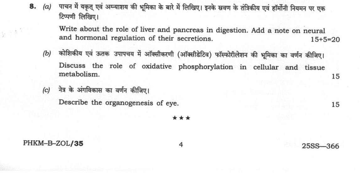
(a) Write about the role of liver and pancreas in digestion. Add a note on neural and hormonal regulation of their secretions…
Write about the role of liver and pancreas in digestion. Add a note on neural and hormonal regulation of their secretions. (15+5=20 marks)
Discuss the role of oxidative phosphorylation in cellular and tissue metabolism. 15 marks
Describe the organogenesis of eye. 15 marks
हिंदी में प्रश्न पढ़ें
पाचन में यकृत एवं अग्न्याशय की भूमिका के बारे में लिखिए। इनके स्रवण के तंत्रिकीय एवं हार्मोनी नियमन पर एक टिप्पणी लिखिए। (15+5=20 अंक)
कोशिकीय एवं उत्तक उपापचय में ऑक्सीकरणी (ऑक्सीडेटिव) फॉस्फोरिलेशन की भूमिका का वर्णन कीजिए। (15 अंक)
नेत्र के अंगविकास का वर्णन कीजिए। (15 अंक)
Model answer
Written by UPSC Answer Check against this question's marking rubric, to the expected length. UPSC does not publish answers for Mains — this is one way to score well, not an official key.
Digestive and Regulatory Roles of the Liver and Pancreas
The liver and pancreas serve as the primary accessory organs coordinating chemical digestion and metabolic homeostasis. The liver synthesizes bile, composed of bile salts (sodium glycocholate and taurocholate), phospholipids, cholesterol, and bile pigments. Bile salts lower surface tension to emulsify dietary triglycerides into micro-droplets, expanding the surface area for enzymatic attack and forming mixed micelles for lipid absorption. The liver also conjugates and excretes bilirubin, the end-product of erythrocyte heme catabolism.
The exocrine pancreas secretes an alkaline pancreatic juice containing bicarbonate ions (HCO₃⁻) to neutralize acidic gastric chyme, alongside vital digestive hydrolases: endopeptidases (trypsinogen, chymotrypsinogen), exopeptidases (procarboxypeptidases), pancreatic alpha-amylase for carbohydrate breakdown, pancreatic lipase for triacylglycerol hydrolysis, and nucleases. Enteropeptidase on the duodenal brush border activates trypsinogen into trypsin, triggering an enzymatic activation cascade. Concurrently, the endocrine pancreas (islets of Langerhans) releases insulin from β-cells and glucagon from α-cells, dynamically regulating postprandial and fasting systemic glucose flux.
Secretion is tightly regulated by coordinated neural and hormonal axes. Neural control operates via cephalic and gastric phases through the vagus nerve (cranial nerve X); cholinergic vago-vagal reflexes stimulate initial basal enzymatic release and gallbladder tone. Hormonal control dominates during the intestinal phase: acidic chyme (pH < 4.5) triggers duodenal S-cells to release secretin, which stimulates ductal secretion of water and bicarbonate. Fat and amino acids stimulate duodenal I-cells to secrete cholecystokinin (CCK), inducing pancreatic acinar enzyme release, gallbladder contraction, and relaxation of the sphincter of Oddi. Gastric inhibitory polypeptide (GIP) and gastrin augment endocrine priming and gastrointestinal motility.
Clinically, hepatic pathology manifests as jaundice, categorized into pre-hepatic (hemolytic), intra-hepatic (hepatocellular damage common in viral hepatitis), and post-hepatic (obstructive gallstones or biliary atresia). In India, chronic pancreatitis associated with malnutrition or alcohol abuse leads to exocrine pancreatic insufficiency, resulting in severe steatorrhea, weight loss, and secondary fat-soluble vitamin malabsorption.
Oxidative Phosphorylation in Cellular and Tissue Metabolism
Oxidative phosphorylation is the terminal biochemical pathway coupling nutrient oxidation to adenosine triphosphate (ATP) synthesis within the inner mitochondrial membrane. Electrons derived from glycolysis, the Krebs cycle, and β-oxidation enter the electron transport chain (ETC) via NADH at Complex I (NADH:ubiquinone oxidoreductase) and FADH₂ at Complex II (succinate dehydrogenase). Electrons transfer sequentially through ubiquinone, Complex III (cytochrome bc₁), cytochrome c, and Complex IV (cytochrome c oxidase), where molecular oxygen is reduced to water.
According to Mitchell’s chemiosmotic hypothesis, electron transit powers proton pumping across Complexes I, III, and IV into the intermembrane space, generating an electrochemical proton gradient. Protons return to the mitochondrial matrix down this gradient through the F_0F₁-ATP synthase complex, driving the rotary catalysis of ADP phosphorylation to ATP, yielding P/O ratios of approximately 2.5 for NADH and 1.5 for FADH₂. Cytosolic reducing equivalents cross into the matrix via the malate-aspartate shuttle (predominant in high-demand cardiac and liver tissue, yielding maximal ATP) or the glycerol-3-phosphate shuttle (predominant in skeletal muscle and brain for rapid regeneration).
Tissue-specific adaptations address divergent metabolic roles. Highly oxidative tissues, such as the myocardium and central nervous system, maintain dense cristae and high baseline oxidative phosphorylation to meet non-negotiable ATP demands, whereas skeletal muscle transitions between oxidative phosphorylation in slow-twitch Type I fibers and anaerobic glycolysis in fast-twitch Type II fibers. In brown adipose tissue (BAT), uncoupling protein-1 (UCP-1 or thermogenin) short-circuits the proton gradient, dissipating proton motive energy as heat for non-shivering thermogenesis rather than generating ATP.
Organogenesis and Developmental Genetics of the Eye
Eye organogenesis is a classic model of reciprocal embryonic induction involving neuroectoderm, surface ectoderm, and periocular mesenchyme. Development initiates during early neurulation with the lateral evagination of the diencephalon to form paired optic vesicles. As an optic vesicle contacts the overlying surface ectoderm, it induces the ectoderm to thicken into a lens placode while itself invaginating to form the double-walled optic cup.
The lens placode invaginates to form the lens vesicle, which detaches from the surface ectoderm; its posterior cells elongate into primary lens fibers, establishing optic transparency. Concurrently, the inner layer of the optic cup differentiates into the stratified neural retina, generating photoreceptors (rods and cones), bipolar cells, horizontal cells, amacrine cells, and retinal ganglion cells. The outer layer of the optic cup becomes the single-layered retinal pigment epithelium (RPE). Mesenchymal and cranial neural crest cells condense around the optic cup to form the vascular choroid, the protective fibrous sclera, and the anterior cornea, while the hyaloid vascular system transiently supplies the developing lens before regressing to form the clear vitreous humor.
This morphological sequence is directed by a conserved genetic hierarchy governed by the master regulator Pax6, alongside homeobox transcription factors Rx and Six3, which establish eye field specification and regulate tissue competence. The ventral groove of the optic cup, termed the optic fissure, must close precisely around the hyaloid vessels; failure of this closure produces congenital coloboma. In the Indian public health landscape, identifying structural developmental defects alongside preventable nutritional insults, notably Vitamin A deficiency causing xerophthalmia and keratomalacia, forms an operational priority under the National Programme for Control of Blindness and Visual Impairment (NPCBVI).
Integrating early embryological screening with targeted metabolic and nutritional interventions represents the most effective strategy to prevent congenital malformations, optimize tissue bioenergetics, and mitigate chronic gastrointestinal and sensory pathologies across populations.
What "Discuss" is asking you to do
Lay the issue out from more than one side — how it arose, what is claimed for it, what is held against it, and where it now stands. UPSC attaches discuss to broad topics with several live dimensions, so coverage of the dimensions earns more than the strength of your opinion.
Structure that answers it
Set the issue up → the case as it is made → the case against → the dimension both sides leave out → where the balance now lies
Where marks are lost
Listing facts with no thread between them, or arguing one side throughout and calling it a discussion.
How this answer will be evaluated
Approach
Framework: Zoology Paper 2: Define > Structure/Mechanism > Diagram > Example. (a) explain: definition/context > points in order > small example > short close | (b) discuss: intro > 3-4 dimensions > example > balanced close | (c) describe: define > structure or process in order > labelled diagram > significance Full marks: Precise mechanisms, labelled diagrams, and clear linkage of structure to function.
Key points expected
- Liver: bile synthesis, storage, and role in lipid digestion
- Pancreas: exocrine enzymes (amylase, lipase, trypsin) and bicarbonate secretion
- Neural regulation: vagal stimulation of pancreatic secretion
- Hormonal regulation: CCK and secretin roles in pancreatic/bile secretion
- Definition of oxidative phosphorylation and its location (mitochondria)
- Electron transport chain (ETC) complexes and proton gradient generation
- Chemiosmotic coupling and ATP synthase mechanism
- Link to cellular energy (ATP) production for tissue function
Evaluation rubric
Each sub-part is marked on its own, against the marks and word limit printed on the paper.
- (a) Digestive roles of liver/pancreas plus neural/hormonal regulation of secretions. 20 marks
explain— definition/context → points in order → small example → short close
Must cover
- Liver: bile synthesis, storage, and role in lipid digestion
- Pancreas: exocrine enzymes (amylase, lipase, trypsin) and bicarbonate secretion
- Neural regulation: vagal stimulation of pancreatic secretion
- Hormonal regulation: CCK and secretin roles in pancreatic/bile secretion
Loses marks
- Confusing endocrine (insulin) with exocrine pancreatic function
- Omitting the specific role of bile in fat digestion
- Listing hormones without explaining their target organs
Earns more
- Mechanism of bile salt micelle formation
- Zymogen activation cascade (enterokinase to trypsin)
- Feedback loops involving enteroendocrine cells
Extra mark
- Labelled diagram of hepatopancreatic duct system
- Specific mention of sphincter of Oddi function
- (b) Role of oxidative phosphorylation in cellular and tissue metabolism. 15 marks
discuss— intro → 3-4 dimensions → example → balanced close
Must cover
- Definition of oxidative phosphorylation and its location (mitochondria)
- Electron transport chain (ETC) complexes and proton gradient generation
- Chemiosmotic coupling and ATP synthase mechanism
- Link to cellular energy (ATP) production for tissue function
Loses marks
- Confusing glycolysis with oxidative phosphorylation
- Failing to link the process to ATP synthesis
- Omitting the role of the proton gradient
Earns more
- Specific mention of NADH and FADH2 as electron donors
- Role of oxygen as the final electron acceptor
- Connection to aerobic respiration efficiency
Extra mark
- Labelled diagram of the mitochondrial ETC
- Mention of uncoupling proteins or thermogenesis
- (c) Organogenesis of the eye. 15 marks
describe— define → structure or process in order → labelled diagram → significance
Must cover
- Induction of the optic vesicle from the diencephalon
- Formation of the optic cup and lens vesicle
- Differentiation of the retina (neural and pigment layers)
- Development of the cornea and lens
Loses marks
- Omitting the induction process (optic vesicle to cup)
- Confusing the origin of the lens (ectoderm) with the retina (neuroectoderm)
- Failing to describe the formation of the optic nerve
Earns more
- Role of the surface ectoderm in lens formation
- Formation of the optic stalk and nerve
- Development of the vitreous body
Extra mark
- Labelled diagram of the developing eye stages
- Mention of specific embryonic days/weeks for key stages
Practice this exact question
Write your answer and it is marked point by point against the model answer above — what you covered, what you missed, what you got wrong.
Evaluate my answer →More from Zoology 2024 Paper II
- Q5 Write on the following in about 150 words each: (a) Functions of immunoglobulins (10 mark…
- Q6 (a) Explain the structure of nephron and its role in urine formation. Add a note on hormo…
- Q7 (a) Describe the process of spermatogenesis. Add a note on the role of Golgi bodies in th…
- Q8 (a) Write about the role of liver and pancreas in digestion. Add a note on neural and hor…