Paper I — Q3
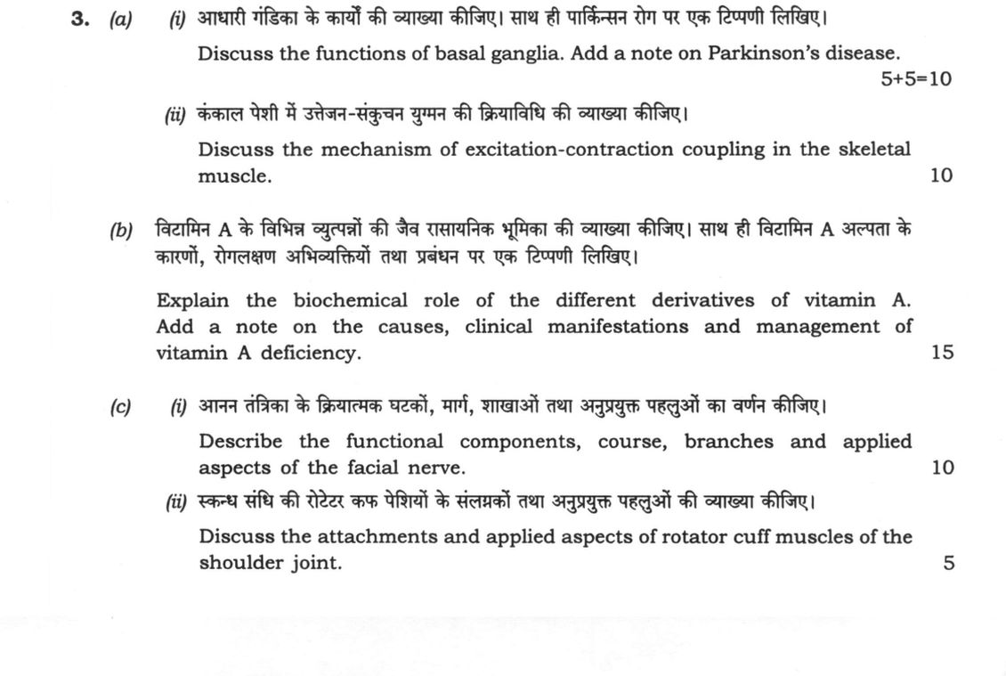
(a) (i) Discuss the functions of basal ganglia. Add a note on Parkinson's disease. (5+5=10 marks) (ii) Discuss the mechanism of…
Discuss the functions of basal ganglia. Add a note on Parkinson's disease. (5+5=10 marks)
Discuss the mechanism of excitation-contraction coupling in the skeletal muscle. 10 marks
Explain the biochemical role of the different derivatives of vitamin A. Add a note on the causes, clinical manifestations and management of vitamin A deficiency. 15 marks
Describe the functional components, course, branches and applied aspects of the facial nerve. 10 marks
Discuss the attachments and applied aspects of rotator cuff muscles of the shoulder joint. 5 marks
हिंदी में प्रश्न पढ़ें
आधारी गांडिका के कार्यों की व्याख्या कीजिए। साथ ही पार्किनसन रोग पर एक टिप्पणी लिखिए। (5+5=10)
कंकाल पेशी में उत्तेजन-संकुचन युग्मन की क्रियाविधि की व्याख्या कीजिए। 10 marks
विटामिन A के विभिन्न व्युत्पन्नों की जैव रासायनिक भूमिका की व्याख्या कीजिए। साथ ही विटामिन A अल्पता के कारणों, रोगलक्षण अभिव्यक्तियों तथा प्रबंधन पर एक टिप्पणी लिखिए। 15 marks
आनन तंत्रिका के क्रियात्मक घटकों, मार्ग, शाखाओं तथा अनुप्रयुक्त पहलुओं का वर्णन कीजिए। 10 marks
स्कंध संधि की रोटर कफ पेशियों के संलग्नकों तथा अनुप्रयुक्त पहलुओं की व्याख्या कीजिए। 5 marks
Model answer
Written by UPSC Answer Check against this question's marking rubric, to the expected length. UPSC does not publish answers for Mains — this is one way to score well, not an official key.
Basal Ganglia and Parkinson’s Disease The basal ganglia (caudate, putamen, globus pallidus, substantia nigra, and subthalamic nucleus) regulate voluntary motor execution, motor learning, and cognitive-emotional loops. Motor control relies on a balance between two striatocortical circuits: the direct pathway (striatal D1 receptors projecting to GPi/SNpr) which disinhibits the thalamus to initiate movement, and the indirect pathway (striatal D2 receptors via GPe and STN) which increases GPi/SNpr output to inhibit unnecessary motor programs. Parkinson’s disease arises from the progressive loss of dopaminergic neurons in the substantia nigra pars compacta (SNpc), marked histopathologically by Lewy bodies containing aggregated α-synuclein. The resulting loss of striatal dopamine shifts the pathway balance toward excessive indirect pathway hyperactivity. Clinically, this manifests as the classic "TRAP" tetrad: resting pill-rolling Tremor, lead-pipe or cogwheel Rigidity, Akinesia or bradykinesia, and Postural instability.
Excitation-Contraction Coupling in Skeletal Muscle Excitation-contraction coupling translates a sarcolemmal action potential into mechanical tension. Depolarization propagates along the transverse tubules (T-tubules), activating voltage-gated L-type calcium channels, the dihydropyridine receptors (DHPR). Mechanical coupling between DHPR and ryanodine receptors (RyR1) on the sarcoplasmic reticulum (SR) terminal cisternae triggers massive Ca²⁺ release into the sarcoplasm. Cytoplasmic Ca²⁺ binds to troponin C on the thin filament, inducing a conformational shift in the troponin-tropomyosin complex that exposes the myosin-binding sites on actin. Cross-bridge cycling ensues: energized myosin heads bind actin, release inorganic phosphate, perform the power stroke, bind ATP to detach, and hydrolyze ATP to re-cock. Relaxation occurs when Ca²⁺ is actively pumped back into the SR lumen by the sarcoplasmic/endoplasmic reticulum Ca²⁺-ATPase (SERCA) pump and buffered by calsequestrin.
Biochemical Role and Deficiency of Vitamin A Vitamin A derivatives perform distinct functions: Retinal (11-cis-retinal) combines with opsin in retinal rod cells to form rhodopsin. Light isomerizes 11-cis-retinal to all-trans-retinal, triggering a transducin-mediated cGMP phosphodiesterase cascade that hyperpolarizes the photoreceptor, enabling scotopic (dim-light) vision (Wald's visual cycle). Retinoic acid functions as a ligand for nuclear receptors (RAR and RXR), acting as a transcription factor to maintain normal epithelial differentiation, mucus secretion, and immune competence. Retinol represents the primary transport (bound to retinol-binding protein) and storage form (retinyl palmitate in hepatic stellate cells), essential for reproductive function.
Deficiency results from primary dietary lack (addressed in India via the National Prophylaxis Programme against Nutritional Blindness and ICDS), fat malabsorption syndromes (celiac disease, cholestasis), or advanced liver dysfunction. Clinical manifestations progress according to WHO staging: night blindness (nyctalopia), conjunctival xerosis, Bitot’s spots (keratinized triangular conjunctival plaques), corneal xerosis, and irreversible keratomalacia (liquefactive corneal necrosis leading to blindness). Management involves oral retinol therapy (200,000 IU for age >1 year on days 0, 1, and 14; intramuscular formulations in malabsorption) alongside prophylactic biannual mega-dose Vitamin A Supplementation (VAS).
Facial Nerve and Rotator Cuff Anatomy The facial nerve (cranial nerve VII) contains four functional components: Special Visceral Efferent (SVE to muscles of facial expression, stapedius, stylohyoid, posterior digastric), Special Sensory (SVA taste from anterior two-thirds of tongue via chorda tympani), General Visceral Efferent (GVE parasympathetic to lacrimal, sublingual, and submandibular glands via greater petrosal and chorda tympani), and General Somatic Afferent (GSA sensation from the external acoustic meatus). Its course traverses the cerebellopontine angle, enters the internal acoustic meatus, travels the Z-shaped facial canal (giving off the greater petrosal nerve, nerve to stapedius, and chorda tympani), and exits the stylomastoid foramen. It then pierces the parotid gland to divide into five terminal motor branches: temporal, zygomatic, buccal, marginal mandibular, and cervical. Lower motor neuron lesion (Bell’s palsy) causes ipsilateral flaccid paralysis of facial muscles, assessed clinically via the House-Brackmann scale; inability to close the eye (lagophthalmos) risks exposure keratitis, mandating immediate corneal lubrication and tarsorrhaphy if severe.
The rotator cuff consists of the SITS muscles which dynamic-stabilize the glenohumeral joint: Supraspinatus (supraspinous fossa to upper facet of greater tubercle; initiates abduction 0–15°). Infraspinatus (infraspinous fossa to middle facet of greater tubercle; lateral rotator). Teres minor (lateral scapular border to lower facet of greater tubercle; lateral rotator). Subscapularis (subscapular fossa to lesser tubercle; medial rotator). Clinically, subacromial impingement of the supraspinatus tendon beneath the coracoacromial arch causes a "painful arc" between 60° and 120° of active abduction, frequently progressing to degenerative cuff tears in manual laborers and overhead athletes.
Addressing these neurological, musculoskeletal, and metabolic conditions requires early diagnostic recognition (such as testing the painful arc or identifying Bitot's spots) combined with evidence-based interventions like dopamine replacement therapy, surgical tendon repair, and sustained national public-health nutritional supplementation.
What "Discuss" is asking you to do
Lay the issue out from more than one side — how it arose, what is claimed for it, what is held against it, and where it now stands. UPSC attaches discuss to broad topics with several live dimensions, so coverage of the dimensions earns more than the strength of your opinion.
Structure that answers it
Set the issue up → the case as it is made → the case against → the dimension both sides leave out → where the balance now lies
Where marks are lost
Listing facts with no thread between them, or arguing one side throughout and calling it a discussion.
How this answer will be evaluated
Approach
Framework: Medical Science, Paper 1. (a(i)) discuss: intro > 3-4 dimensions > example > balanced close | (a(ii)) discuss: intro > 3-4 dimensions > example > balanced close | (b) explain: definition/context > points in order > small example > short close | (c(i)) describe: define > structure or process in order > labelled diagram > significance | (c(ii)) discuss: intro > 3-4 dimensions > example > balanced close Full marks: Comprehensive, accurate, and well-structured answers with all required components and applied aspects.
Key points expected
- Motor control functions (initiation, modulation)
- Non-motor functions (cognitive, emotional)
- Pathophysiology of Parkinson's (dopamine loss)
- Clinical features of Parkinson's disease
- Action potential propagation along T-tubules
- Calcium release from sarcoplasmic reticulum
- Troponin-tropomyosin interaction
- Cross-bridge cycling and ATP hydrolysis
Evaluation rubric
Each sub-part is marked on its own, against the marks and word limit printed on the paper.
- (a(i)) Functions of basal ganglia and a note on Parkinson's disease. 10 marks
discuss— intro → 3-4 dimensions → example → balanced close
Must cover
- Motor control functions (initiation, modulation)
- Non-motor functions (cognitive, emotional)
- Pathophysiology of Parkinson's (dopamine loss)
- Clinical features of Parkinson's disease
Loses marks
- Listing functions without mechanism
- Confusing Parkinson's with other movement disorders
Earns more
- Mention of basal ganglia nuclei (caudate, putamen)
- Direct and indirect pathways
- Levodopa therapy
Extra mark
- Mention of specific neurotransmitters (GABA, acetylcholine)
- (a(ii)) Mechanism of excitation-contraction coupling in skeletal muscle. 10 marks
discuss— intro → 3-4 dimensions → example → balanced close
Must cover
- Action potential propagation along T-tubules
- Calcium release from sarcoplasmic reticulum
- Troponin-tropomyosin interaction
- Cross-bridge cycling and ATP hydrolysis
Loses marks
- Omitting the role of calcium
- Confusing cardiac and skeletal muscle mechanisms
Earns more
- Role of dihydropyridine receptors (DHR)
- Ryanodine receptors (RyR)
- Sliding filament theory
Extra mark
- Diagram of the coupling mechanism
- (b) Biochemical role of vitamin A derivatives and deficiency management. 15 marks
explain— definition/context → points in order → small example → short close
Must cover
- Role of retinal in vision (rhodopsin)
- Role of retinoic acid in gene expression
- Causes of vitamin A deficiency
- Clinical manifestations (night blindness, xerophthalmia)
Loses marks
- Listing symptoms without biochemical mechanism
- Management without priority or context
Earns more
- Management strategies (supplementation)
- Prevention programs (National health programme)
- Role in immune function
Extra mark
- Mention of specific WHO guidelines
- (c(i)) Functional components, course, branches, and applied aspects of the facial nerve. 10 marks
describe— define → structure or process in order → labelled diagram → significance
Must cover
- Functional components (motor, sensory, parasympathetic)
- Course through the facial canal
- Major branches (temporal, zygomatic, buccal, marginal, cervical)
- Applied aspects (Bell's palsy, parotid surgery)
Loses marks
- Omitting the parasympathetic component
- Confusing facial nerve with trigeminal nerve branches
Earns more
- Labelled diagram of the facial nerve
- Mention of chorda tympani
- Clinical tests for facial nerve function
Extra mark
- Mention of specific surgical landmarks
- (c(ii)) Attachments and applied aspects of rotator cuff muscles. 5 marks
discuss— intro → 3-4 dimensions → example → balanced close
Must cover
- Attachments of the four rotator cuff muscles
- Function of the rotator cuff
- Applied aspects (rotator cuff tears, impingement syndrome)
Loses marks
- Omitting the function of the rotator cuff
- Confusing rotator cuff muscles with deltoid
Earns more
- Mention of specific muscles (supraspinatus, infraspinatus, teres minor, subscapularis)
- Clinical tests for rotator cuff integrity
Extra mark
- Mention of specific surgical repair techniques
Practice this exact question
Write your answer and it is marked point by point against the model answer above — what you covered, what you missed, what you got wrong.
Evaluate my answer →More from Medical Science 2022 Paper I
- Q1 (a) Discuss the stomach under the following headings: (i) Peritoneal relations (ii) Blood…
- Q2 (a) Describe the anatomy of uterus under the following headings: (i) Gross features (ii)…
- Q3 (a) (i) Discuss the functions of basal ganglia. Add a note on Parkinson's disease. (5+5=1…
- Q4 (a) (i) Explain the mechanism of iron absorption in the intestine. What are the factors r…
- Q5 (a) Describe the molecular mechanisms of carcinogenesis of breast carcinoma. Describe the…
- Q6 (a) (i) Describe in brief the pathogenesis and histopathological features of rheumatic he…