Paper I — Q1
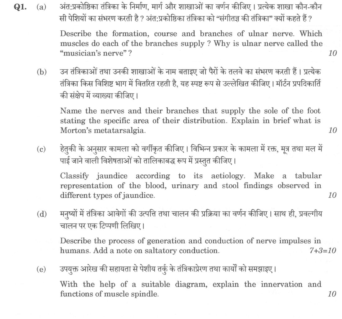
(a) Describe the formation, course and branches of ulnar nerve. Which muscles do each of the branches supply ? Why is ulnar nerve…
Describe the formation, course and branches of ulnar nerve. Which muscles do each of the branches supply ? Why is ulnar nerve called the "musician's nerve" ? 10 marks
Name the nerves and their branches that supply the sole of the foot stating the specific area of their distribution. Explain in brief what is Morton's metatarsalgia. 10 marks
Classify jaundice according to its aetiology. Make a tabular representation of the blood, urinary and stool findings observed in different types of jaundice. 10 marks
Describe the process of generation and conduction of nerve impulses in humans. Add a note on saltatory conduction. 7+3=10 marks
With the help of a suitable diagram, explain the innervation and functions of muscle spindle. 10 marks
हिंदी में प्रश्न पढ़ें
अंतःप्रकोष्ठिका तंत्रिका के निर्माण, मार्ग और शाखाओं का वर्णन कीजिए। प्रत्येक शाखा कौन-कौन सी पेशियों का संभरण करती है ? अंतःप्रकोष्ठिका तंत्रिका को "संगीतज्ञ की तंत्रिका" क्यों कहते हैं ? 10 अंक
उन तंत्रिकाओं तथा उनकी शाखाओं के नाम बताइए जो पैरों के तलवे का संभरण करती हैं। प्रत्येक तंत्रिका किस विशिष्ट भाग में वितरित रहती है, यह स्पष्ट रूप से उल्लेखित कीजिए। मॉर्टन प्रपदिकाति की संक्षेप में व्याख्या कीजिए। 10 अंक
हेतुकी के अनुसार कामला को वर्गीकृत कीजिए। विभिन्न प्रकार के कामला में रक्त, मूत्र तथा मल में पाई जाने वाली विशेषताओं को तालिकाबद्ध रूप में प्रस्तुत कीजिए। 10 अंक
मनुष्यों में तंत्रिका आवेगों की उत्पत्ति तथा चालन की प्रक्रिया का वर्णन कीजिए। साथ ही, प्रवल्गीय चालन पर एक टिप्पणी लिखिए। 7+3=10 अंक
उपयुक्त आरेख की सहायता से पेशीय तर्कु के तंत्रिकाप्रेरण तथा कार्यों को समझाइए। 10 अंक
Model answer
Written by UPSC Answer Check against this question's marking rubric, to the expected length. UPSC does not publish answers for Mains — this is one way to score well, not an official key.
(a) Ulnar nerve. The ulnar nerve arises from the medial cord of the brachial plexus (C8–T1). In the axilla it lies posterior to the axillary artery; in the arm it descends in the medial bicipital groove, medial to the brachial artery. At the elbow it passes posterior to the medial epicondyle through the cubital tunnel, between the medial epicondyle and olecranon. In the forearm it lies between the heads of flexor carpi ulnaris and on flexor digitorum profundus, medial to the ulnar artery. It gives muscular branches to flexor carpi ulnaris and the medial half of flexor digitorum profundus. The palmar cutaneous branch arises proximal to the wrist, passes superficial to the flexor retinaculum and supplies the skin of the medial palm. The dorsal cutaneous branch arises 5–8 cm proximal to the wrist, passes posterior to the wrist, deep to the extensor retinaculum, and supplies the dorsum of the hand and medial 1½ fingers. The nerve enters Guyon’s canal at the wrist and divides into superficial and deep branches. The superficial branch supplies palmaris brevis and palmar skin of the medial 1½ fingers. The deep branch supplies the hypothenar muscles (abductor digiti minimi, flexor digiti minimi brevis, opponens digiti minimi), the third and fourth lumbricals, all interossei, adductor pollicis and the deep head of flexor pollicis brevis. It is called the “musician’s nerve” because these intrinsic hand muscles control fine finger movements, abduction, adduction and opposition required for playing piano, guitar and violin; compression or injury impairs precision grip and dexterity.
(b) Plantar innervation and Morton’s metatarsalgia. The sole is supplied by branches of the tibial nerve and, for the lateral heel, by the sural nerve. Behind the medial malleolus the tibial nerve gives medial calcaneal branches to the skin of the medial heel, then divides into medial and lateral plantar nerves. The medial plantar nerve supplies the skin of the medial sole and medial 3½ toes, and the muscles abductor hallucis, flexor digitorum brevis, flexor hallucis brevis and the first lumbrical. The lateral plantar nerve supplies the skin of the lateral sole and lateral 1½ toes, and the muscles quadratus plantae, abductor digiti minimi, flexor digiti minimi brevis, adductor hallucis, interossei and the second to fourth lumbricals. The sural nerve supplies the lateral heel. Morton’s metatarsalgia is an interdigital neuroma, usually between the third and fourth metatarsal heads, where the common digital nerve is compressed by the intermetatarsal ligament; it causes burning pain, paraesthesia and a clicking sensation in the forefoot, worsened by tight shoes.
(c) Jaundice. Jaundice is classified aetiologically as pre-hepatic, hepatic and post-hepatic. Pre-hepatic jaundice results from increased bilirubin production, e.g. haemolytic anaemia, malaria, G6PD deficiency or transfusion reaction. Hepatic jaundice results from hepatocellular injury, e.g. viral hepatitis A or E, alcoholic liver disease, cirrhosis, drug-induced injury, Wilson’s disease or alpha-1 antitrypsin deficiency. Post-hepatic jaundice results from obstruction to bile flow, e.g. choledocholithiasis, cholangiocarcinoma, carcinoma of head of pancreas, biliary stricture or Ascaris in the common bile duct.
| Type | Blood | Urine | Stool | |---|---|---|---| | Pre-hepatic | Unconjugated bilirubin increased; conjugated normal | Bilirubin absent; urobilinogen increased | Normal or dark | | Hepatic | Unconjugated and conjugated bilirubin increased | Bilirubin present; urobilinogen increased or variable | Normal to pale | | Post-hepatic | Conjugated bilirubin increased; unconjugated normal | Bilirubin present; urobilinogen decreased or absent | Acholic/clay-coloured |
(d) Nerve impulse. The resting axonal membrane is about –70 mV, maintained by the Na+/K+ ATPase and selective K+ permeability. A stimulus that reaches threshold opens voltage-gated Na+ channels; Na+ influx depolarises the membrane to about +30 mV. Na+ channels then inactivate and voltage-gated K+ channels open, so K+ efflux repolarises the membrane. A brief hyperpolarisation follows because K+ channels close slowly. The absolute refractory period prevents a second impulse, and the relative refractory period requires a stronger stimulus. Conduction occurs because local currents depolarise the adjacent membrane, regenerating the action potential along the axon; it is all-or-none and non-decremental. In saltatory conduction, myelin sheaths formed by Schwann cells in the peripheral nervous system or oligodendrocytes in the central nervous system insulate the axon, leaving nodes of Ranvier rich in voltage-gated Na+ channels. The action potential regenerates only at the nodes and appears to jump from node to node, making conduction faster and more energy-efficient because fewer ions cross the membrane.
(e) Muscle spindle. The muscle spindle is a fusiform proprioceptor within skeletal muscle. It contains intrafusal fibres—nuclear bag and nuclear chain fibres—surrounded by a capsule, with extrafusal fibres outside. Sensory endings are Ia annulospiral endings on the bag and chain fibres and II flower-spray endings mainly on chain fibres. Efferent supply is by gamma motor neurons to the intrafusal fibres and alpha motor neurons to the extrafusal fibres.
Diagram: ``text Ia annulospiral afferent \ \ Tendon [ ( nuclear bag fibre ) ] Tendon / Tendon [ ( nuclear chain fibre ) ] Tendon / II flower-spray afferent gamma motor neuron → intrafusal fibres alpha motor neuron → extrafusal fibres ``
The Ia afferents enter the spinal cord and form monosynaptic excitatory connections with alpha motor neurons of the same muscle, producing the stretch reflex; they also inhibit antagonist motoneurons. The II afferents contribute to proprioception and slower stretch responses. The gamma motor neurons adjust intrafusal fibre tension, maintaining spindle sensitivity during muscle contraction; this alpha-gamma co-activation allows the spindle to monitor muscle length throughout movement. Thus the spindle functions as a length detector, initiates the stretch reflex, provides proprioceptive information, and helps coordinate posture and fine motor control. These structures and processes show the integrated basis of motor control, sensory function and clinical jaundice.
What "Describe" is asking you to do
Give a full, ordered account of the thing named — its parts, stages or mechanism — in the sequence in which it actually exists or occurs. Most describe questions come from the science optionals, where the marks sit in correct technical detail and, where the stem says so, a labelled diagram.
Structure that answers it
One-line identification of the subject → the parts or stages in their real order, each with its defining detail → labelled diagram where the subject is structural → closing line on function or significance
Where marks are lost
Loose general prose where the examiner is ticking named parts, correct terminology and their sequence; and in the General Studies papers, turning to evaluation before the description is finished.
How this answer will be evaluated
Approach
Framework: Medical Science, Paper 1. (a) describe: define > structure or process in order > labelled diagram > significance | (b) describe: define > structure or process in order > labelled diagram > significance | (c) compare: paired headings or table > key differences > significance > conclusion | (d) describe: define > structure or process in order > labelled diagram > significance | (e) explain: definition/context > points in order > small example > short close Full marks: Accurate anatomical/physiological details, clear diagrams, and precise tabular data.
Key points expected
- Formation from medial cord of brachial plexus
- Course through cubital fossa and Guyon's canal
- Muscle supply of each branch (e.g., FCU, medial FDP)
- Explanation of 'musician's nerve' (intrinsic hand muscle loss)
- Tibial nerve branches (medial/lateral plantar nerves)
- Specific distribution areas of plantar nerves
- Definition of Morton's metatarsalgia (interdigital neuroma)
- Location of Morton's neuroma (3rd web space)
Evaluation rubric
Each sub-part is marked on its own, against the marks and word limit printed on the paper.
- (a) Formation, course, branches, muscle supply, and 'musician's nerve' rationale of ulnar nerve. 10 marks
describe— define → structure or process in order → labelled diagram → significance
Must cover
- Formation from medial cord of brachial plexus
- Course through cubital fossa and Guyon's canal
- Muscle supply of each branch (e.g., FCU, medial FDP)
- Explanation of 'musician's nerve' (intrinsic hand muscle loss)
Loses marks
- Omitting specific muscle names for branches
- Confusing ulnar with median nerve course
Earns more
- Labelled diagram of ulnar nerve course
- Mention of sensory distribution (medial 1.5 digits)
Extra mark
- Clinical test for ulnar nerve palsy (Froment's sign)
- (b) Nerves/branches of sole of foot with distribution areas and definition of Morton's metatarsalgia. 10 marks
describe— define → structure or process in order → labelled diagram → significance
Must cover
- Tibial nerve branches (medial/lateral plantar nerves)
- Specific distribution areas of plantar nerves
- Definition of Morton's metatarsalgia (interdigital neuroma)
- Location of Morton's neuroma (3rd web space)
Loses marks
- Listing nerves without specific distribution areas
- Confusing Morton's metatarsalgia with general metatarsalgia
Earns more
- Mention of sural nerve contribution
- Diagram of plantar nerve distribution
Extra mark
- Mention of Morton's test (compression sign)
- (c) Aetiological classification of jaundice and tabular comparison of blood, urine, and stool findings. 10 marks
compare— paired headings or table → key differences → significance → conclusion
Must cover
- Classification: Pre-hepatic, Hepatic, Post-hepatic
- Table comparing blood bilirubin (unconjugated vs conjugated)
- Table comparing urine findings (urobilinogen/bilirubin)
- Table comparing stool findings (acholic vs normal)
Loses marks
- Missing the tabular format for findings
- Confusing conjugated vs unconjugated bilirubin in urine
Earns more
- Specific examples for each aetiological type
- Mention of specific enzymes (e.g., ALP, Transaminases)
Extra mark
- Mention of specific causes like Gilbert's syndrome
- (d) Generation and conduction of nerve impulses plus a note on saltatory conduction. 10 marks
describe— define → structure or process in order → labelled diagram → significance
Must cover
- Resting membrane potential and action potential phases
- Ion movements (Na+ influx, K+ efflux)
- Mechanism of saltatory conduction (node to node)
- Role of myelin sheath in saltatory conduction
Loses marks
- Omitting the specific note on saltatory conduction
- Incorrect ion movement description
Earns more
- Diagram of action potential curve
- Mention of refractory period
Extra mark
- Comparison of myelinated vs unmyelinated conduction speed
- (e) Innervation and functions of muscle spindle with a suitable diagram. 10 marks
explain— definition/context → points in order → small example → short close
Must cover
- Structure of muscle spindle (intrafusal fibers)
- Innervation: Ia afferent, II afferent, gamma efferent
- Function: Stretch reflex (monosynaptic)
- Role in muscle tone and proprioception
Loses marks
- Missing the required diagram
- Confusing muscle spindle with Golgi tendon organ
Earns more
- Labelled diagram of muscle spindle innervation
- Mention of Golgi tendon organ for contrast
Extra mark
- Explanation of gamma loop mechanism
Practice this exact question
Write your answer and it is marked point by point against the model answer above — what you covered, what you missed, what you got wrong.
Evaluate my answer →More from Medical Science 2025 Paper I
- Q1 (a) Describe the formation, course and branches of ulnar nerve. Which muscles do each of…
- Q2 (a) Using a well labelled diagram, trace the taste pathway from circumvallate papillae of…
- Q3 (a) A 36-year-old cyclist suffered pelvic injury while riding into a pothole on the road.…
- Q4 (a) (i) A 55-year-old lady visits surgery OPD with a three month old history of a painles…