Paper II — Q2
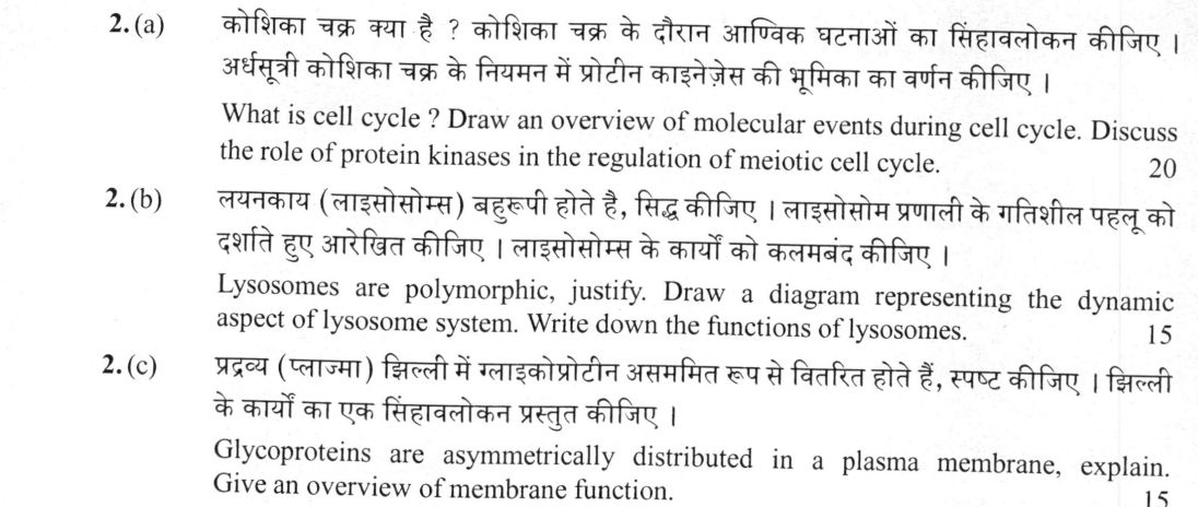
(a) What is cell cycle ? Draw an overview of molecular events during cell cycle. Discuss the role of protein kinases in the…
What is cell cycle ? Draw an overview of molecular events during cell cycle. Discuss the role of protein kinases in the regulation of meiotic cell cycle. 20 marks
Lysosomes are polymorphic, justify. Draw a diagram representing the dynamic aspect of lysosome system. Write down the functions of lysosomes. 15 marks
Glycoproteins are asymmetrically distributed in a plasma membrane, explain. Give an overview of membrane function. 15 marks
हिंदी में प्रश्न पढ़ें
कोशिका चक्र क्या है ? कोशिका चक्र के दौरान आणविक घटनाओं का सिंहावलोकन कीजिए । अर्धसूत्री कोशिका चक्र के नियमन में प्रोटीन काइनेजेस की भूमिका का वर्णन कीजिए । 20
लयनकाय (लाइसोसोम्स) बहुरूपी होते हैं, सिद्ध कीजिए । लाइसोसोम प्रणाली के गतिशील पहलू को दर्शाते हुए आरेखित कीजिए । लाइसोसोम्स के कार्यों को कलमबद्ध कीजिए । 15
प्रद्रव्य (प्लाज्मा) झिल्ली में ग्लाइकोप्रोटीन असममित रूप से वितरित होते हैं, स्पष्ट कीजिए । झिल्ली के कार्यों का एक सिंहावलोकन प्रस्तुत कीजिए । 15
Model answer
Written by UPSC Answer Check against this question's marking rubric, to the expected length. UPSC does not publish answers for Mains — this is one way to score well, not an official key.
(a) Cell Cycle and Meiotic Kinase Regulation
The cell cycle is an ordered series of events through which a cell duplicates its genome and contents, culminating in cell division. It comprises Interphase (G₁, S, G₂) and M phase (mitosis/meiosis), governed by surveillance checkpoints (G₁/S, G₂/M, and the Spindle Assembly Checkpoint).
Molecular Events and Cyclin-CDK Oscillation:
`` [G1 Phase: Cyclin D-CDK4/6] ---> Rb phosphorylation / E2F release │ [G1/S Checkpoint: Cyclin E-CDK2] ---> Pre-replication complex firing │ [S Phase: Cyclin A-CDK2] ---> DNA replication │ [G2/M Checkpoint: Cyclin A/B-CDK1] ---> Wee1 vs. Cdc25 regulation │ [M Phase: Active MPF (Cyclin B-CDK1)] ---> Spindle assembly & APC/C activation ``
Role of Protein Kinases in Meiotic Cell Cycle: Meiotic progression is driven by the Maturation-Promoting Factor (MPF), a heterodimer of CDK1 (Cdc2 kinase) and Cyclin B. In prophase I arrest (diplotene/dictyate stage), CDK1 is kept inactive by dual phosphorylation at Thr14 and Tyr15 by the inhibitory kinase Wee1/Myt1.
Re-entry into meiosis I involves the activation of Cdc25 phosphatase alongside activating phosphorylation by CDK-activating kinase (CAK) at Thr161, which overrides Wee1 inhibition. Active MPF phosphorylates lamins (nuclear envelope breakdown), condensins (chromosome condensation), and histone H3.
Unlike mitosis, exit from meiosis I requires partial inactivation of MPF driven by the Anaphase-Promoting Complex/Cyclosome (APC/C^Cdc20), degrading Cyclin B without permitting DNA replication between Meiosis I and II (S-phase suppression). Entry into Meiosis II requires rapid re-accumulation of Cyclin B-CDK1. Metaphase II arrest (in vertebrates) is maintained by the Cytostatic Factor (CSF/Emi2 pathway) inhibiting APC/C, until fertilization-induced calcium influx activates calcineurin and CaMKII, destroying CSF, degrading Cyclin B via APC/C, and driving anaphase II completion.
(b) Polymorphism and Dynamic System of Lysosomes
Lysosomes exhibit marked structural and functional polymorphism due to varying enzymatic content, stage of maturation, and the substrate being digested.
Forms and Pathways: Primary lysosomes are nascent vesicles budded from the *trans*-Golgi network containing inactive acid hydrolases. Secondary lysosomes arise through fusion with substrate-bearing vesicles: heterophagosomes (digesting extracellular material via heterophagy), autophagosomes (degrading damaged organelles via autophagy), and crinosomes (degrading excess secretory granules via crinophagy). Residual bodies contain undigested debris destined for exocytosis or accumulation.
Dynamic Lysosome System: `` Extracellular Matter ──> Endocytosis ──> Early Endosome ──> Late Endosome ┐ │ (Fusion) Golgi (Hydrolases) ────> Primary Lysosome ────────────────────────────────┴──> Secondary Lysosome │ Damaged Organelles ────> Autophagosome ────────────────────────────────────────────┤ (Digestion) ▼ Exocytosis / Lipofuscin <── Residual Body <────────────────────────────────────────┘ ``
Functions of Lysosomes:
- Intracellular Digestion: Hydrolysis of proteins, nucleic acids, lipids, and carbohydrates via ~50 acid hydrolases (pH ~4.5–5.0).
- Autophagy and Organelle Turnover: Recycling cellular components during starvation and clearing protein aggregates.
- Apoptosis: Lysosomal membrane permeabilization releases cathepsins into cytosol, activating intrinsic apoptotic pathways.
- Extracellular Remodeling: Osteoclasts secrete lysosomal enzymes (e.g., Cathepsin K) into the sub-osteoclastic space for bone resorption.
- Pathological Relevance: Defects in specific hydrolases cause Lysosomal Storage Diseases, such as ganglioside accumulation in Tay-Sachs disease and glucocerebroside buildup in Gaucher disease.
(c) Glycoprotein Asymmetry and Plasma Membrane Function
Asymmetry of Glycoproteins: Plasma membrane glycoproteins display absolute structural asymmetry: oligosaccharide moieties are localized exclusively on the non-cytosolic (extracellular) leaflet. This asymmetry is established cotranslationally and post-translationally within the lumens of the rough endoplasmic reticulum and Golgi apparatus. Because polar carbohydrate groups cannot cross the hydrophobic core of the bilayer, and phospholipid translocators (flippases/scramblases) do not translocate bulky glycosylated proteins, the luminal orientation is maintained during vesicle fusion with the plasma membrane. These external sugar residues form the glycocalyx (cell coat), essential for cell-cell recognition (e.g., selectins in leukocyte homing), receptor protection, and histocompatibility (MHC complexes).
Overview of Membrane Functions:
- Selective Permeability and Transport: Mediates passive movement (simple diffusion, facilitated transport via ion channels and aquaporins) and primary/secondary active transport against electrochemical gradients (e.g., Na⁺/K⁺ ATPase).
- Signal Transduction: Transmits extracellular cues via G-protein-coupled receptors (GPCRs), receptor tyrosine kinases (RTKs), and ligand-gated channels to regulate intracellular cascades.
- Cell Adhesion and Junctions: Mediates tissue coherence via cadherins, integrins, occludins, and claudins in desmosomes, tight junctions, and hemidesmosomes.
- Membrane Potential Maintenance: Generates resting membrane potentials and action potentials through asymmetric ion distribution, underlying neuromuscular excitability.
Coordinated regulation of cell cycle kinases, endomembrane dynamics, and asymmetric membrane architecture is fundamental to maintaining cellular homeostasis and multicellular organization.
What "Discuss" is asking you to do
Lay the issue out from more than one side — how it arose, what is claimed for it, what is held against it, and where it now stands. UPSC attaches discuss to broad topics with several live dimensions, so coverage of the dimensions earns more than the strength of your opinion.
Structure that answers it
Set the issue up → the case as it is made → the case against → the dimension both sides leave out → where the balance now lies
Where marks are lost
Listing facts with no thread between them, or arguing one side throughout and calling it a discussion.
How this answer will be evaluated
Approach
Framework: UPSC Zoology Paper 2. (a) discuss: intro > 3-4 dimensions > example > balanced close | (b) justify: claim > 3-4 reasons > evidence > conclusion | (c) explain: definition/context > points in order > small example > short close Full marks: Precise definitions, labelled diagrams, specific molecular mechanisms, and named examples.
Key points expected
- Define cell cycle and its phases (G1, S, G2, M)
- Draw overview of molecular events (DNA replication, spindle formation)
- Discuss role of protein kinases (CDKs, Cyclins) in meiosis
- Explain specific kinases like MPF (Maturation Promoting Factor)
- Justify polymorphism via different shapes/sizes (primary, secondary, residual)
- Draw diagram of dynamic aspect (autophagy, heterophagy, exocytosis)
- List functions (digestion, autophagy, apoptosis)
- Mention hydrolytic enzymes (acid hydrolases)
Evaluation rubric
Each sub-part is marked on its own, against the marks and word limit printed on the paper.
- (a) Define cell cycle, draw molecular events, and discuss protein kinase regulation in meiosis. 20 marks
discuss— intro → 3-4 dimensions → example → balanced close
Must cover
- Define cell cycle and its phases (G1, S, G2, M)
- Draw overview of molecular events (DNA replication, spindle formation)
- Discuss role of protein kinases (CDKs, Cyclins) in meiosis
- Explain specific kinases like MPF (Maturation Promoting Factor)
Loses marks
- Confusing mitotic and meiotic specific events
- Diagram without labels for molecular components
- Failing to link kinases to specific cell cycle transitions
Earns more
- Mention specific cyclin-CDK complexes (e.g., Cyclin B-CDK1)
- Reference to checkpoint controls (G1/S, G2/M)
- Mention of phosphatase role in kinase regulation
Extra mark
- Reference to specific model organism (e.g., Xenopus oocytes)
- Mention of specific molecular inhibitors (e.g., roscovitine)
- (b) Justify lysosome polymorphism, draw dynamic system diagram, and list functions. 15 marks
justify— claim → 3-4 reasons → evidence → conclusion
Must cover
- Justify polymorphism via different shapes/sizes (primary, secondary, residual)
- Draw diagram of dynamic aspect (autophagy, heterophagy, exocytosis)
- List functions (digestion, autophagy, apoptosis)
- Mention hydrolytic enzymes (acid hydrolases)
Loses marks
- Diagram missing the dynamic flow (formation to degradation)
- Failing to distinguish primary vs secondary lysosomes
- Listing functions without linking to mechanism
Earns more
- Mention of lysosomal storage diseases (e.g., Tay-Sachs)
- Reference to pH dependence of enzymes
- Mention of phagosome-lysosome fusion
Extra mark
- Reference to specific enzyme (e.g., acid phosphatase)
- Mention of specific disease (e.g., Gaucher's disease)
- (c) Explain asymmetric distribution of glycoproteins and give overview of membrane function. 15 marks
explain— definition/context → points in order → small example → short close
Must cover
- Explain asymmetric distribution (glycocalyx on extracellular side)
- Mention role of Golgi apparatus in glycosylation
- Give overview of membrane functions (barrier, transport, signaling)
- Mention specific membrane proteins (integral, peripheral)
Loses marks
- Failing to explain why distribution is asymmetric
- Confusing glycoproteins with glycolipids
- Listing functions without linking to structure
Earns more
- Reference to fluid mosaic model (Singer & Nicolson)
- Mention of specific transport mechanisms (active, passive)
- Reference to cell-cell recognition via glycoproteins
Extra mark
- Mention of specific glycoprotein (e.g., integrins)
- Reference to specific membrane function (e.g., endocytosis)
Practice this exact question
Write your answer and it is marked point by point against the model answer above — what you covered, what you missed, what you got wrong.
Evaluate my answer →More from Zoology 2021 Paper II
- Q1 Write your answer in about 150 words for each of the following : 10×5=50 (a) Describe the…
- Q2 (a) What is cell cycle ? Draw an overview of molecular events during cell cycle. Discuss…
- Q3 (a) What is chromosome mutation ? Describe various types of polyploidy with suitable exam…
- Q4 (a) What is isolation ? Describe the major isolating mechanisms which lead to speciation.…
- Q5 Write your answer in about 150 words for each of the following : 10×5=50 (a) Fatty acids…