Paper II — Q7
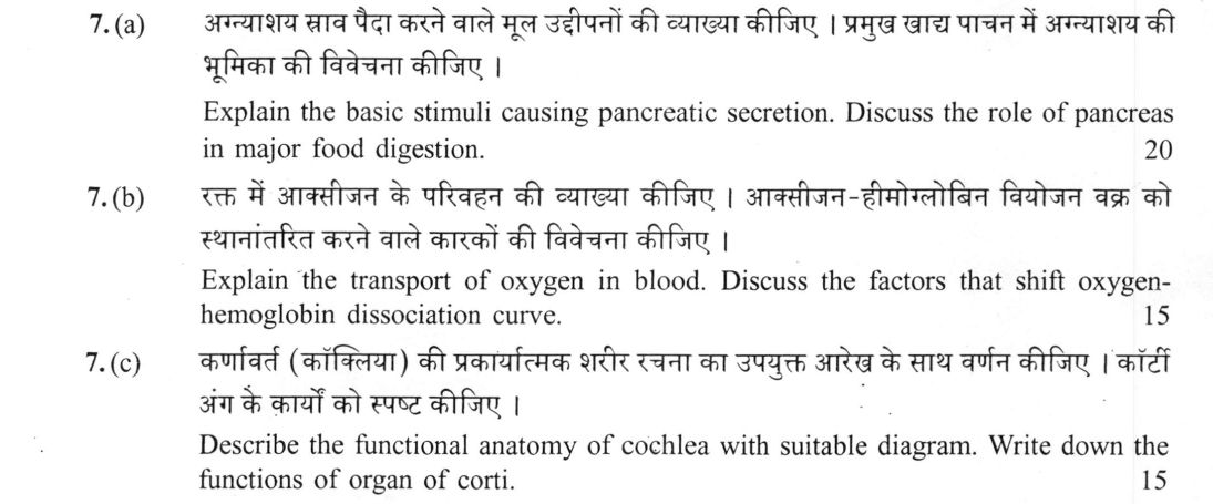
(a) Explain the basic stimuli causing pancreatic secretion. Discuss the role of pancreas in major food digestion. 20 (b) Explain…
Explain the basic stimuli causing pancreatic secretion. Discuss the role of pancreas in major food digestion. 20 marks
Explain the transport of oxygen in blood. Discuss the factors that shift oxygen-hemoglobin dissociation curve. 15 marks
Describe the functional anatomy of cochlea with suitable diagram. Write down the functions of organ of corti. 15 marks
हिंदी में प्रश्न पढ़ें
अग्न्याशय स्राव पैदा करने वाले मूल उद्दीपनों की व्याख्या कीजिए । प्रमुख खाद्य पाचन में अग्न्याशय की भूमिका की विवेचना कीजिए । 20
रक्त में आक्सीजन के परिवहन की व्याख्या कीजिए । आक्सीजन-हीमोग्लोबिन विमोजन वक्र को स्थानान्तरित करने वाले कारकों की विवेचना कीजिए । 15
कर्णवर्त (कॉक्लिया) की प्रकार्यात्मक शरीर रचना का उपयुक्त आरेख के साथ वर्णन कीजिए । कॉर्टी अंग के कार्यों को स्पष्ट कीजिए । 15
Model answer
Written by UPSC Answer Check against this question's marking rubric, to the expected length. UPSC does not publish answers for Mains — this is one way to score well, not an official key.
Pancreatic secretion and digestion. Pancreatic secretion is initiated when food enters the duodenum. In the neural phase, distension and vagal afferents excite vagal efferents to acinar cells, causing early enzyme-rich secretion. The vagal phase is brief and prepares the pancreas before food arrives, while hormonal secretion is the main phase during digestion. In the hormonal phase, acid and peptides in the duodenum stimulate S cells to release secretin, which acts on duct cells to secrete bicarbonate-rich fluid, neutralising gastric acid and providing an alkaline pH for enzymes. Fatty acids and amino acids stimulate I cells to release cholecystokinin, which mainly stimulates acinar cells to secrete digestive enzymes and also promotes gallbladder contraction. Acinar cells package amylase, lipase, trypsinogen, chymotrypsinogen and procarboxypeptidases; duct cells add bicarbonate and water. In digestion, pancreatic α-amylase hydrolyses internal α-1,4 glycosidic bonds of gelatinised starch, producing maltose, maltotriose and α-limit dextrins; it does not act effectively on raw starch granules. Proteolytic zymogens are activated by enterokinase: trypsinogen becomes trypsin, which activates chymotrypsinogen and procarboxypeptidases; trypsin, chymotrypsin and carboxypeptidases hydrolyse peptide bonds, releasing amino acids and small peptides. Pancreatic lipase, with colipase and bile salts, hydrolyses triglycerides to fatty acids and 2-monoglycerides, enabling micelle formation and absorption. Thus the pancreas provides the alkaline medium and enzymes for carbohydrate, protein and lipid digestion.
Oxygen transport and dissociation curve. Oxygen is carried in blood mainly as oxyhaemoglobin, about 98.5%, and only about 1.5% as dissolved plasma oxygen, the dissolved fraction determining blood pO2. Haemoglobin is a tetramer with four heme groups; binding of one O2 molecule increases affinity of the remaining sites, giving cooperative binding represented as Hb + 4O2 ⇌ Hb(O2)4. The oxygen-haemoglobin dissociation curve is sigmoidal, allowing efficient loading at pulmonary pO2 and unloading at tissue pO2. A right shift, caused by increased pCO2, decreased pH (Bohr effect), increased temperature and increased 2,3-BPG, lowers haemoglobin affinity and promotes O2 unloading in metabolically active tissues. During exercise, increased tissue pCO2, H+ and temperature shift the curve right, facilitating unloading. A left shift, caused by decreased pCO2, increased pH, lower temperature or lower 2,3-BPG, increases affinity and promotes loading in the lungs. 2,3-BPG binds deoxyhaemoglobin and stabilises the low-affinity state; fetal haemoglobin has reduced 2,3-BPG binding affinity because of γ-globin chains, not absent binding sites, so it retains higher O2 affinity for placental transfer. Thus the curve shifts according to tissue needs, matching oxygen supply to metabolism.
Cochlear anatomy and organ of Corti. A suitable diagram shows the cochlea as a bony spiral with three fluid-filled chambers: scala vestibuli and scala tympani containing perilymph, separated by the cochlear duct or scala media containing endolymph. The vestibular membrane separates scala vestibuli from scala media; the basilar membrane separates scala media from scala tympani. The helicotrema at the apex connects scala vestibuli and scala tympani. The round window at the base bulges to dissipate pressure. Sound vibrations pass from the stapes footplate to the oval window, generating pressure waves in perilymph that travel along the basilar membrane. The basilar membrane is stiff and narrow at the base and broad and flexible at the apex, so high frequencies produce maximal displacement near the base and low frequencies near the apex, providing frequency discrimination. On the basilar membrane lies the organ of Corti, with inner and outer hair cells, supporting cells and the overlying tectorial membrane. When the basilar membrane moves, stereocilia of hair cells shear against the tectorial membrane, opening mechanosensitive ion channels; depolarisation releases neurotransmitter onto auditory nerve fibres. Inner hair cells are principal sensory receptors encoding sound intensity and frequency, while outer hair cells amplify and sharpen the basilar membrane response. Together, the cochlea converts mechanical sound into neural signals and separates frequencies for hearing.
What "Explain" is asking you to do
Make the working of something clear — what sets it off, what follows from what, and what it produces. Explain is the Commission's mechanism word: it dominates the technical papers and the “explain why” stems, where the marks sit in the causal chain and not in the label.
Structure that answers it
State what it is → the initiating condition → the chain of cause, step by step → an instance where it plays out → what the chain produces
Where marks are lost
Describing what something looks like instead of why it works that way. Naming the stages without linking them reads as description too.
How this answer will be evaluated
Approach
Framework: Zoology Paper 2: Define > Structure/Mechanism > Diagram > Example. (a) explain: definition/context > points in order > small example > short close | (b) explain: definition/context > points in order > small example > short close | (c) describe: define > structure or process in order > labelled diagram > significance Full marks: Precise mechanisms, labelled diagrams, and specific physiological links.
Key points expected
- Neural (vagal) and hormonal (CCK, secretin) stimuli
- Mechanism of enzyme and bicarbonate secretion
- Role in protein, lipid, and carbohydrate digestion
- Specific enzymes (trypsin, lipase, amylase) named
- Mechanism of O2 binding to hemoglobin
- Factors shifting the curve (Bohr effect, pH, CO2, temp)
- Explanation of right vs. left shift significance
- Labelled dissociation curve diagram
Evaluation rubric
Each sub-part is marked on its own, against the marks and word limit printed on the paper.
- (a) Mechanism of pancreatic secretion stimuli and digestive roles. 20 marks
explain— definition/context → points in order → small example → short close
Must cover
- Neural (vagal) and hormonal (CCK, secretin) stimuli
- Mechanism of enzyme and bicarbonate secretion
- Role in protein, lipid, and carbohydrate digestion
- Specific enzymes (trypsin, lipase, amylase) named
Loses marks
- Confusing gastric and pancreatic enzymes
- Omitting the role of bicarbonate in neutralization
- Vague description of 'digestion' without specific substrates
Earns more
- Distinction between exocrine and endocrine functions
- Feedback loops in secretion regulation
- Clinical relevance (e.g., pancreatitis)
Extra mark
- Specific receptor types (e.g., CCK-A)
- Quantitative data on enzyme activity
- (b) Oxygen transport mechanisms and dissociation curve shifts. 15 marks
explain— definition/context → points in order → small example → short close
Must cover
- Mechanism of O2 binding to hemoglobin
- Factors shifting the curve (Bohr effect, pH, CO2, temp)
- Explanation of right vs. left shift significance
- Labelled dissociation curve diagram
Loses marks
- Drawing curve without axes or labels
- Confusing oxygenation with carbon dioxide transport
- Failing to link factors to physiological state
Earns more
- Mention of 2,3-DPG (2,3-BPG) role
- Distinction between fetal and adult hemoglobin
- Link to altitude or diving physiology
Extra mark
- Specific p50 values for different conditions
- Mention of myoglobin as a secondary store
- (c) Functional anatomy of cochlea and Organ of Corti. 15 marks
describe— define → structure or process in order → labelled diagram → significance
Must cover
- Labelled diagram of cochlear cross-section
- Structure of Organ of Corti (hair cells, tectorial membrane)
- Mechanism of transduction (stereocilia bending)
- Tonal topography (base vs. apex)
Loses marks
- Diagram without labels (specifically hair cells)
- Confusing cochlea with semicircular canals
- Omitting the function of the tectorial membrane
Earns more
- Mention of endolymph vs. perilymph ionic composition
- Role of basilar membrane resonance
- Link to auditory nerve transmission
Extra mark
- Specific hair cell types (inner vs. outer)
- Mention of otoacoustic emissions
Practice this exact question
Write your answer and it is marked point by point against the model answer above — what you covered, what you missed, what you got wrong.
Evaluate my answer →More from Zoology 2021 Paper II
- Q4 (a) What is isolation ? Describe the major isolating mechanisms which lead to speciation.…
- Q5 Write your answer in about 150 words for each of the following : 10×5=50 (a) Fatty acids…
- Q6 (a) What are peptide hormones ? With the help of schematic diagram, discuss the epinephri…
- Q7 (a) Explain the basic stimuli causing pancreatic secretion. Discuss the role of pancreas…
- Q8 (a) What are morphogens ? Describe the cellular differentiation during morphogenesis. 20…