Paper I — Q7
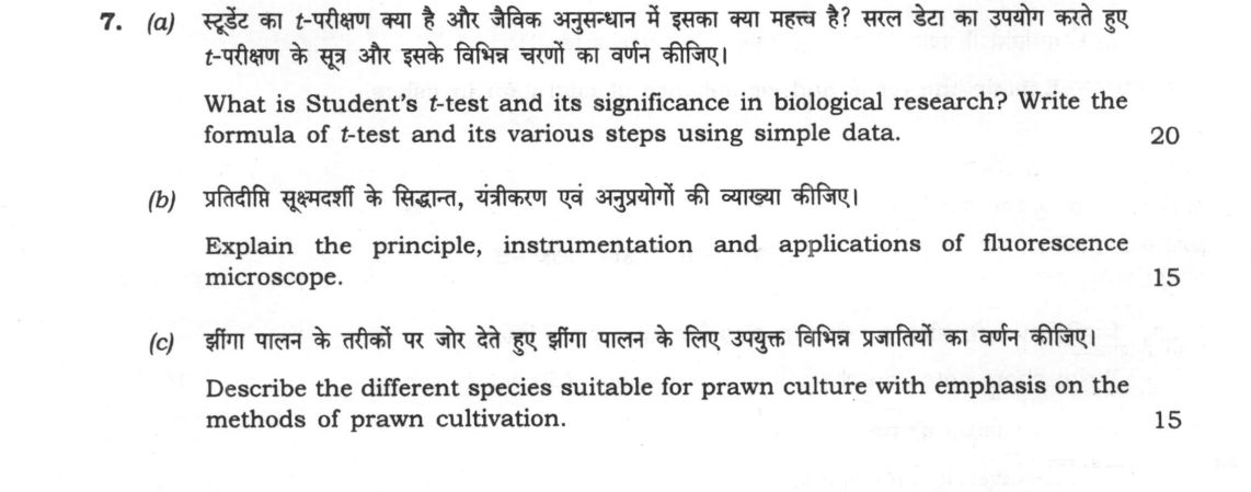
(a) What is Student's t-test and its significance in biological research? Write the formula of t-test and its various steps using…
What is Student's t-test and its significance in biological research? Write the formula of t-test and its various steps using simple data. 20 marks
Explain the principle, instrumentation and applications of fluorescence microscope. 15 marks
Describe the different species suitable for prawn culture with emphasis on the methods of prawn cultivation. 15 marks
हिंदी में प्रश्न पढ़ें
स्टूडेंट का t-परीक्षण क्या है और जैविक अनुसंधान में इसका क्या महत्व है? सरल डेटा का उपयोग करते हुए t-परीक्षण के सूत्र और इसके विभिन्न चरणों का वर्णन कीजिए। 20
प्रतिदीप्ति सूक्ष्मदर्शी के सिद्धांत, यंत्रीकरण एवं अनुप्रयोगों की व्याख्या कीजिए। 15
झींगा पालन के तरीकों पर जोर देते हुए झींगा पालन के लिए उपयुक्त विभिन्न प्रजातियों का वर्णन कीजिए। 15
Model answer
Written by UPSC Answer Check against this question's marking rubric, to the expected length. UPSC does not publish answers for Mains — this is one way to score well, not an official key.
Student’s t-Test in Biological Research
Student’s t-test is a parametric statistical test developed by W.S. Gosset to determine whether the difference between the means of two groups is statistically significant when sample sizes are small (n < 30) and population variance is unknown. Its validity rests on assumptions of a normal distribution of the variable, homogeneity of variance (homoscedasticity), and independent, random sampling. In biological research, it is indispensable for testing null hypotheses (H₀) versus alternative hypotheses (H₁) in experimental setups—such as evaluating drug efficacy, toxicological impacts, or physiological variations between control and treatment groups—while controlling Type I (α) and Type II (β) errors.
For two independent samples, the test statistic is calculated as: t = (x̄₁ - x̄₂)/(√(sₚ² (1/(n₁) + 1/(n₂)))) where the pooled variance is sₚ² = (Σ (x₁ - x̄₁)² + Σ (x₂ - x̄₂)²)/(n₁ + n₂ - 2) and degrees of freedom df = n₁ + n₂ - 2.
Step-by-step calculation on fish weight gain (g) under Control (A: 4, 5, 6) and Supplemented Diet (B: 7, 8, 9):
- Compute sample sizes and means: n₁ = 3, x̄₁ = 5; n₂ = 3, x̄₂ = 8.
- Compute sum of squared deviations: Σ(x₁ - x̄₁)² = (4-5)² + 0² + (6-5)² = 2; Σ(x₂ - x̄₂)² = (7-8)² + 0² + (9-8)² = 2.
- Calculate pooled variance: sₚ² = (2 + 2) / (3 + 3 - 2) = 1.0.
- Calculate standard error of difference: SE = √(1.0 × (1/3 + 1/3)) = √0.667 ≈ 0.816.
- Compute calculated t: t = |5 - 8| / 0.816 = 3.68.
- Decision: At df = 4 and α = 0.05, the critical value from the t-table is t_crit = 2.776. Because calculated t (3.68) > t_crit (2.776), H₀ is rejected, confirming that the supplemented diet produces a statistically significant increase in growth.
Fluorescence Microscopy
Principle: Fluorescence microscopy relies on fluorophores absorbing high-energy, shorter-wavelength light (excitation) and emitting lower-energy, longer-wavelength light (emission). The difference between excitation and emission maxima is the Stokes shift. Unlike bright-field and phase-contrast microscopy, which rely on light absorption or phase shifts through whole specimens, fluorescence microscopy selectively visualizes specific labeled cellular components against a completely dark background, generating exceptional contrast and sensitivity.
Instrumentation: In an epifluorescence setup, a light source (mercury/xenon arc lamp or laser) passes light through an excitation filter, which transmits only the specific excitation wavelength. A dichroic mirror reflects this short-wavelength beam through the objective lens onto the specimen. The fluorophores emit longer-wavelength light, which passes back through the objective and transmits across the dichroic mirror. An emission (barrier) filter removes stray excitation light, transmitting pure fluorescence to the detector (CCD camera or oculars).
Applications: Key uses include immunofluorescence for subcellular antigen localization, Fluorescence In Situ Hybridization (FISH) for gene mapping and chromosomal aberration detection, Green Fluorescent Protein (GFP) tagging to track protein dynamics in live-cell imaging, cancer diagnostic marker detection, and neurobiological neuronal tracing.
Prawn and Shrimp Culture
Suitable Species: In India, commercially cultured species comprise marine and brackishwater penaeids like Penaeus monodon (giant tiger prawn) and Litopenaeus vannamei (Pacific white shrimp, favored for its Specific Pathogen Free status and fast growth), alongside the freshwater palaemonid Macrobrachium rosenbergii (giant freshwater prawn/scampi).
Cultivation Methods:
- Extensive Culture: Practiced in traditional coastal ecosystems (such as pokkali fields in Kerala and bheries in West Bengal) utilizing tidal water exchange, low stocking densities (<5 PL/m²), and natural pond productivity.
- Semi-Intensive Culture: Uses engineered earthen ponds, stocking densities of 10--25 PL/m², pond fertilization, commercial formulated feeds, and regular water exchange.
- Intensive Culture: Employs high stocking densities (>40 PL/m²), closed biosecure systems, formulated pellet diets, and continuous paddlewheel aeration.
Management and Systems: Rearing involves hatchery production of post-larvae (PL), nursery acclimatization, and strict monitoring of dissolved oxygen, salinity, pH, and ammonia. Biosecurity protocols mitigate viral threats like White Spot Syndrome Virus (WSSV) and bacterial Early Mortality Syndrome (EMS). Freshwater scampi is widely cultivated in polyculture with Indian Major Carps. Coastal prawn aquaculture in India is strictly regulated under the Coastal Aquaculture Authority (CAA) and MPEDA guidelines to enforce biosecurity, effluent treatment, and environmental sustainability.
What "Explain" is asking you to do
Make the working of something clear — what sets it off, what follows from what, and what it produces. Explain is the Commission's mechanism word: it dominates the technical papers and the “explain why” stems, where the marks sit in the causal chain and not in the label.
Structure that answers it
State what it is → the initiating condition → the chain of cause, step by step → an instance where it plays out → what the chain produces
Where marks are lost
Describing what something looks like instead of why it works that way. Naming the stages without linking them reads as description too.
How this answer will be evaluated
Approach
Framework: null. (a) explain: definition/context > points in order > small example > short close | (b) explain: definition/context > points in order > small example > short close | (c) describe: define > structure or process in order > labelled diagram > significance Full marks: Precise definitions, correct formulas/calculations, labelled diagrams, and specific named examples.
Key points expected
- Definition of Student's t-test
- Formula for t-value (t = mean diff / SE)
- Step-by-step calculation using provided simple data
- Significance in biological research
- Principle of fluorescence (absorption/emission)
- Instrumentation details (excitation/emission filters)
- Applications in biological research
- Diagram of the microscope setup
Evaluation rubric
Each sub-part is marked on its own, against the marks and word limit printed on the paper.
- (a) Define t-test, state formula, and demonstrate calculation steps using simple data. 20 marks
explain— definition/context → points in order → small example → short close
Must cover
- Definition of Student's t-test
- Formula for t-value (t = mean diff / SE)
- Step-by-step calculation using provided simple data
- Significance in biological research
Loses marks
- Missing formula
- Calculation errors in data steps
- No mention of biological significance
Earns more
- Mention of degrees of freedom
- Comparison with critical t-value
- Distinction between paired and unpaired t-test
Extra mark
- Correct interpretation of p-value
- (b) Explain the working principle, instrumentation, and applications of fluorescence microscopy. 15 marks
explain— definition/context → points in order → small example → short close
Must cover
- Principle of fluorescence (absorption/emission)
- Instrumentation details (excitation/emission filters)
- Applications in biological research
- Diagram of the microscope setup
Loses marks
- Confusing principle with simple light microscopy
- Missing instrumentation details
- No applications listed
Earns more
- Mention of specific fluorochromes
- Comparison with bright-field microscopy
Extra mark
- Mention of confocal microscopy
- (c) Describe species suitable for prawn culture and methods of prawn cultivation. 15 marks
describe— define → structure or process in order → labelled diagram → significance
Must cover
- Named species suitable for culture (e.g., Penaeus monodon)
- Methods of prawn cultivation (pond preparation, stocking)
- Harvesting and post-harvest handling
- Emphasis on cultivation methods as requested
Loses marks
- Using common names only without scientific names
- Neglecting the 'methods' emphasis
- No specific species named
Earns more
- Mention of specific water quality parameters
- Feed management details
Extra mark
- Mention of specific disease management
Practice this exact question
Write your answer and it is marked point by point against the model answer above — what you covered, what you missed, what you got wrong.
Evaluate my answer →More from Zoology 2025 Paper I
- Q4 (a) Draw a well-labelled diagram of testis and ovary in mammals, and discuss the function…
- Q5 Write short notes on the following in about 150 words each: 10×5=50 (a) Biodiversity hot…
- Q6 (a) Define air pollution. Explain the types, sources, consequences and control measures o…
- Q7 (a) What is Student's t-test and its significance in biological research? Write the formu…
- Q8 (a) What do you mean by infectious and communicable diseases? Mention the causative agent…