Paper II — Q5
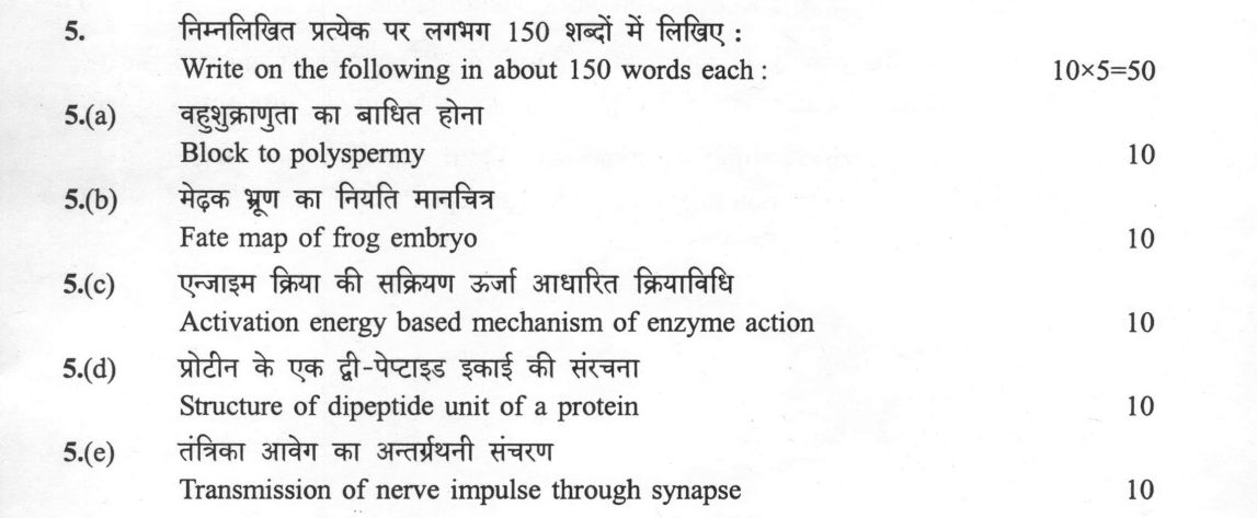
Write on the following in about 150 words each : 10×5=50 (a) Block to polyspermy (10 marks) (b) Fate map of frog embryo (10…
Write on the following in about 150 words each : 10×5=50
Block to polyspermy 10 marks
Fate map of frog embryo 10 marks
Activation energy based mechanism of enzyme action 10 marks
Structure of dipeptide unit of a protein 10 marks
Transmission of nerve impulse through synapse 10 marks
हिंदी में प्रश्न पढ़ें
निम्नलिखित प्रत्येक पर लगभग 150 शब्दों में लिखिए : 10×5=50
बहुशुक्राणुता का बाधित होना (10 अंक)
मेंढक भ्रूण का नियति मानचित्र (10 अंक)
एन्जाइम क्रिया की सक्रियण ऊर्जा आधारित क्रियाविधि (10 अंक)
प्रोटीन के एक डाई-पेप्टाइड इकाई की संरचना (10 अंक)
तंत्रिका आवेग का अंतर्प्रेरणी संचरण (10 अंक)
Model answer
Written by UPSC Answer Check against this question's marking rubric, to the 150-word length. UPSC does not publish answers for Mains — this is one way to score well, not an official key.
Block to polyspermy. Polyspermy is prevented by two sequential blocks. The fast electrical block begins when sperm entry changes the oocyte membrane potential. In sea urchins this is a large, transient depolarization, often described from about −70 mV to positive values, due to Na+ influx; in Xenopus the resting potential is about −30 mV and the fertilization potential is smaller, so the electrical block is weaker. The slow permanent block follows cortical granule exocytosis. Granule contents modify and harden the zona pellucida (or vitelline layer in amphibians), forming a fertilization envelope that prevents further sperm binding or penetration. In mammals, the zona reaction is the principal block, with little or no fast electrical block; in amphibians the slow cortical reaction is decisive. Thus the fast block gives immediate protection, while the slow block provides a durable, species-specific barrier in most eggs, ensuring monospermy and normal embryonic development.
Fate map of frog embryo. The frog blastula fate map shows which surface regions give rise to which tissues. The animal pole, especially the animal cap, is presumptive ectoderm; the dorsal-animal hemisphere forms neural ectoderm and the neural tube. The sub-equatorial marginal zone is presumptive mesoderm: the dorsal and dorsal-lateral marginal zone gives paraxial mesoderm, including somites, and axial structures; the lateral marginal zone gives lateral plate mesoderm; the ventral marginal zone gives ventral mesoderm. The vegetal pole is presumptive endoderm, forming gut and associated organs. Vogt’s vital staining and transplantation experiments established these lineages by following labelled cells. The dorsal lip of the blastopore, within the dorsal marginal zone, acts as the organizer, inducing neural tissue and establishing the dorsal-ventral and anteroposterior axes. Thus the fate map links surface position to germ-layer identity and provides the basis for developmental patterning and organ specification in the embryo.
Activation energy based mechanism of enzyme action. Enzymes increase reaction rates by lowering the activation energy required to reach the transition state. In transition-state theory, the rate depends on the free-energy barrier, ΔG‡; an enzyme binds the transition state more favourably than the substrate, stabilizing it and reducing Ea,catalyzed << Ea,uncatalyzed. In an energy profile, substrate and product free energies remain unchanged, but the catalysed pathway has a lower peak than the uncatalysed pathway. This explains why enzymes accelerate both forward and reverse reactions without changing equilibrium. Michaelis-Menten kinetics captures the functional consequence: V = Vmax[S]/(Km+[S]), where kcat reflects the conversion of enzyme-substrate complex to product. Catalysis mainly increases kcat by lowering the activation barrier, while Km reflects apparent affinity, influenced by binding and catalytic steps. Thus the activation-energy mechanism links molecular binding to measurable enzyme kinetics. It does not consume enzyme and does not alter the reaction’s thermodynamic endpoint.
Structure of dipeptide unit of a protein. A dipeptide, H2N–CHR–CO–NH–CHR–COOH, is formed by condensation between the α-carboxyl group of one amino acid and the α-amino group of another, releasing water and creating a peptide bond. The peptide bond has partial double-bond character because of resonance between the carbonyl oxygen and the amide nitrogen; this makes the C–N bond planar and restricts rotation about it. The ω angle is usually trans, about 180°, placing the two α-carbons on opposite sides, although cis peptide bonds can occur, especially with proline. Rotation is possible mainly about the N–Cα bond, called φ, and the Cα–C bond, called ψ. Steric hindrance among side chains and backbone atoms restricts allowed φ and ψ combinations, as shown in a Ramachandran plot; glycine has greater freedom, while proline is highly restricted. The dipeptide retains a free N-terminus and C-terminus, and its planar peptide backbone is the repeating structural unit of proteins.
Transmission of nerve impulse through synapse. Nerve impulses cross synapses either electrically or chemically. Electrical synapses use gap junctions, allowing direct ion flow; transmission is fast and usually bidirectional. Chemical synapses are one-way because neurotransmitter vesicles are presynaptic and receptors are postsynaptic. When an action potential reaches the presynaptic terminal, voltage-gated Ca2+ channels open, Ca2+ enters, and Ca2+-dependent exocytosis releases neurotransmitter into the synaptic cleft. The transmitter binds postsynaptic receptors; ionotropic receptors open ion channels and produce an EPSP or IPSP depending on the ion conductance, while metabotropic receptors act through second messengers. If depolarization reaches threshold, a postsynaptic action potential is generated. The synaptic delay, about 0.5–1 ms, is the total time from presynaptic action potential to postsynaptic response, arising from Ca2+ entry, vesicle fusion, diffusion, and receptor activation. Termination occurs by reuptake, enzymatic degradation, diffusion, and glial uptake or diffusion. This ensures precise timing and directionality of neural signalling.
What "Describe" is asking you to do
Give a full, ordered account of the thing named — its parts, stages or mechanism — in the sequence in which it actually exists or occurs. Most describe questions come from the science optionals, where the marks sit in correct technical detail and, where the stem says so, a labelled diagram.
Structure that answers it
One-line identification of the subject → the parts or stages in their real order, each with its defining detail → labelled diagram where the subject is structural → closing line on function or significance
Where marks are lost
Loose general prose where the examiner is ticking named parts, correct terminology and their sequence; and in the General Studies papers, turning to evaluation before the description is finished.
How this answer will be evaluated
Approach
Framework: Zoology Paper 2: Define > Structure/Mechanism > Diagram > Example. (a) write short notes: define > 3-4 key features > one example > one-line significance | (b) write short notes: define > 3-4 key features > one example > one-line significance | (c) write short notes: define > 3-4 key features > one example > one-line significance | (d) write short notes: define > 3-4 key features > one example > one-line significance | (e) write short notes: define > 3-4 key features > one example > one-line significance Full marks: Precise mechanism, correct terminology, clear structure, and relevant examples.
Key points expected
- Define polyspermy and its lethal consequence
- Describe the fast block (membrane potential change)
- Describe the slow block (cortical reaction)
- Mention ooplasmic changes (cortical granules)
- Define fate map concept
- Identify dorsal vs ventral blastomere fates
- Identify animal vs vegetal hemisphere fates
- Mention the organizer (Spemann-Mangold)
Evaluation rubric
Each sub-part is marked on its own, against the marks and word limit printed on the paper.
- (a) Mechanism preventing multiple sperm entry into the oocyte. 10 marks · 150 words
write short notes— define → 3-4 key features → one example → one-line significance
Must cover
- Define polyspermy and its lethal consequence
- Describe the fast block (membrane potential change)
- Describe the slow block (cortical reaction)
- Mention ooplasmic changes (cortical granules)
Loses marks
- Confusing fast and slow block mechanisms
- Omitting the role of cortical granules
Earns more
- Mention specific species (e.g., sea urchin)
- Mention role of calcium ion release
- Mention formation of fertilization envelope
Extra mark
- Diagram of cortical granule exocytosis
- (b) Spatial mapping of blastomere fates in frog development. 10 marks · 150 words
write short notes— define → 3-4 key features → one example → one-line significance
Must cover
- Define fate map concept
- Identify dorsal vs ventral blastomere fates
- Identify animal vs vegetal hemisphere fates
- Mention the organizer (Spemann-Mangold)
Loses marks
- Confusing animal and vegetal pole fates
- Failing to mention the organizer
Earns more
- Mention specific species (e.g., Xenopus)
- Mention induction by dorsal lip
- Mention mesoderm induction
Extra mark
- Diagram of frog blastula with fate regions
- (c) How enzymes lower activation energy to catalyze reactions. 10 marks · 150 words
write short notes— define → 3-4 key features → one example → one-line significance
Must cover
- Define activation energy (Ea)
- Explain enzyme as a catalyst
- Describe transition state stabilization
- Mention energy profile diagram concept
Loses marks
- Confusing activation energy with free energy
- Failing to explain the lowering of Ea
Earns more
- Mention specific enzyme (e.g., catalase)
- Mention substrate binding energy
- Mention induced fit model
Extra mark
- Energy profile diagram (Reactants -> TS -> Products)
- (d) Chemical structure of two amino acids linked by a peptide bond. 10 marks · 150 words
write short notes— define → 3-4 key features → one example → one-line significance
Must cover
- Show two amino acid residues
- Show the peptide bond (-CO-NH-)
- Identify N-terminus and C-terminus
- Show the alpha-carbon and side chains
Loses marks
- Incorrect bond connectivity
- Omitting the N and C termini
Earns more
- Mention specific amino acids (e.g., Glycyl-glycine)
- Mention planar nature of peptide bond
- Mention resonance stabilization
Extra mark
- Detailed chemical structure diagram
- (e) Process of signal transmission across the synaptic cleft. 10 marks · 150 words
write short notes— define → 3-4 key features → one example → one-line significance
Must cover
- Describe arrival of action potential
- Describe neurotransmitter release (exocytosis)
- Describe binding to postsynaptic receptors
- Describe postsynaptic potential (EPSP/IPSP)
Loses marks
- Confusing electrical and chemical synapses
- Omitting the role of neurotransmitters
Earns more
- Mention specific neurotransmitter (e.g., Acetylcholine)
- Mention synaptic cleft
- Mention reuptake or degradation
Extra mark
- Diagram of synapse with labeled parts
Practice this exact question
Write your answer and it is marked point by point against the model answer above — what you covered, what you missed, what you got wrong.
Evaluate my answer →More from Zoology 2025 Paper II
- Q2 (a)(i) Describe composition of plasma membrane. (10 marks) (a)(ii) Differentiate between…
- Q3 (a)(i) Describe the synthetic theory of evolution. (10 marks) (a)(ii) Describe common typ…
- Q4 (a) Describe the steps of constructing a recombinant DNA and its cloning. (20 marks) (b)…
- Q5 Write on the following in about 150 words each : 10×5=50 (a) Block to polyspermy (10 mark…
- Q6 (a)(i) Write down the reactions that produce NADH during Krebs cycle. (10 marks) (a)(ii)…
- Q7 (a)(i) Explain sigmoidal nature of oxygen dissociation curve for hemoglobin. (10 marks) (…
- Q8 (a) Describe the stages of fertilization in chick with diagram. (20 marks) (b) Draw struc…