Paper II — Q7
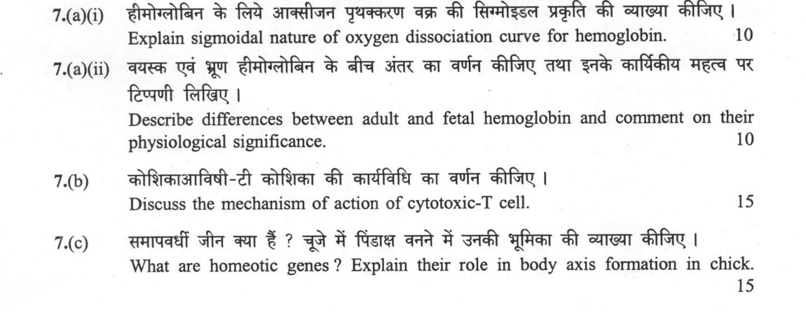
(a)(i) Explain sigmoidal nature of oxygen dissociation curve for hemoglobin. (10 marks) (a)(ii) Describe differences between…
Explain sigmoidal nature of oxygen dissociation curve for hemoglobin. 10 marks
Describe differences between adult and fetal hemoglobin and comment on their physiological significance. 10 marks
Discuss the mechanism of action of cytotoxic-T cell. 15 marks
What are homeotic genes ? Explain their role in body axis formation in chick. 15 marks
हिंदी में प्रश्न पढ़ें
हीमोग्लोबिन के लिए आक्सीजन प्रथक्करण वक्र की सिग्मोइडल प्रकृति की व्याख्या कीजिए । (10 अंक)
वयस्क एवं भ्रूण हीमोग्लोबिन के बीच अंतर का वर्णन कीजिए तथा इनके कार्यिकीय महत्व पर टिप्पणी लिखिए । (10 अंक)
कोशिकाविषी-ट कोशिका की कार्यविधि का वर्णन कीजिए । (15 अंक)
समापवर्धी जीन क्या हैं ? चुजे में पिंडाक्ष बनने में उनकी भूमिका की व्याख्या कीजिए । (15 अंक)
Model answer
Written by UPSC Answer Check against this question's marking rubric, to the expected length. UPSC does not publish answers for Mains — this is one way to score well, not an official key.
The three parts illustrate how molecular interactions produce physiological function, defence and body patterning.
Sigmoidal oxygen dissociation curve. Hemoglobin is an α2β2 tetramer. Binding of the first O2 molecule is weak because deoxygenated Hb is in the low-affinity T-state; this binding induces conformational changes at the α1β2 interfaces that shift the tetramer toward the high-affinity R-state. The R-state increases affinity of the remaining subunits, so O2 binding is cooperative. This produces a sigmoidal curve rather than a hyperbolic one, with a Hill coefficient of about 2.8–3.0. The sigmoid shape is physiologically useful because at high pulmonary pO2 Hb becomes nearly saturated, while at lower tissue pO2 the cooperative transition makes it release O2 efficiently over a narrow pO2 range. The steep middle portion means that a small fall in tissue pO2 causes a large fall in saturation, while pulmonary Hb remains highly saturated. 2,3-BPG binds preferentially to the deoxygenated T-state, stabilizes it and lowers O2 affinity, thereby promoting unloading in metabolically active tissues.
Adult and fetal hemoglobin. Adult HbA is α2β2; fetal HbF is α2γ2. The γ chains differ from β chains in several residues; a common γ-chain polymorphism at position 136 is glycine in Gγ versus alanine in Aγ. More importantly for O2 affinity, γ-chain substitutions reduce 2,3-BPG binding, especially because histidine at β82 is replaced by serine in γ chains. Deoxygenated HbF still adopts a T-state; the reduced 2,3-BPG binding shifts the T–R equilibrium toward R relative to HbA, giving HbF a higher O2 affinity and a lower P50. In the placenta, maternal HbA releases O2 while fetal HbF loads O2 from maternal blood, facilitating transfer to the fetus. This difference in affinity creates a favourable O2 gradient across the placenta. Persistence of HbF is clinically important: in β-thalassaemia it can compensate for defective β chains, and in sickle-cell disease it interferes with HbS polymerization, reducing sickling.
Cytotoxic T-cell mechanism. A cytotoxic T lymphocyte recognizes a specific peptide presented on MHC class I by its TCR, with co-stimulatory signals. This recognition identifies virus-infected or tumour cells. Once engaged, the TCR signal promotes granule mobilization and polarization toward the target cell. Perforin forms pores in the target membrane, allowing granzymes to enter and activate caspases, causing apoptosis. A second pathway is Fas–FasL mediated apoptosis: FasL on the CTL binds Fas on the target, recruiting FADD and caspase-8 to initiate programmed cell death. Apoptosis is non-inflammatory, limiting collateral tissue damage. The CTL survives, proliferates and can form long-lived memory cells that respond rapidly on re-exposure.
Homeotic genes. Homeotic genes are selector genes that assign identity to body segments or regions. In vertebrates they are Hox genes, organized in clusters, and they obey colinearity: gene order on the chromosome corresponds to the order of expression along the anteroposterior axis and to developmental timing. In the chick, Hox genes are expressed in overlapping domains along the neural tube and somites, providing a combinatorial code that specifies cranial, cervical, thoracic and lumbar identities. The anterior-to-posterior expression order ensures each segment receives the correct Hox code. For example, Hoxb-1 is expressed in an anterior hindbrain/cervical domain, while Hoxc-6 is expressed more posteriorly in thoracic regions, helping pattern the neck and trunk. By regulating downstream target genes, Hox proteins determine segment-specific structures such as vertebrae and ribs. Their conservation from Drosophila to vertebrates shows that Hox colinearity is a fundamental mechanism of AP axis formation.
Thus, cooperative Hb conformation, CTL recognition and Hox colinearity each convert molecular interactions into organism-level function: efficient gas exchange, targeted immune killing and correct body patterning.
What "Explain" is asking you to do
Make the working of something clear — what sets it off, what follows from what, and what it produces. Explain is the Commission's mechanism word: it dominates the technical papers and the “explain why” stems, where the marks sit in the causal chain and not in the label.
Structure that answers it
State what it is → the initiating condition → the chain of cause, step by step → an instance where it plays out → what the chain produces
Where marks are lost
Describing what something looks like instead of why it works that way. Naming the stages without linking them reads as description too.
How this answer will be evaluated
Approach
Framework: null. (a(i)) explain: definition/context > points in order > small example > short close | (a(ii)) describe: define > structure or process in order > labelled diagram > significance | (b) discuss: intro > 3-4 dimensions > example > balanced close | (c) explain: definition/context > points in order > small example > short close Full marks: Precise mechanisms, labelled diagrams, specific gene names, and clear physiological links.
Key points expected
- T-state (tense) vs R-state (relaxed) conformational change
- Cooperative binding of O2 to heme groups
- Bohr effect: pH and CO2 influence affinity
- Labelled diagram of sigmoidal curve
- Subunit composition: HbA (α2β2) vs HbF (α2γ2)
- Higher O2 affinity of HbF (left-shifted curve)
- Physiological significance: O2 transfer from mother to fetus
- Mention of 2,3-BPG binding difference
Evaluation rubric
Each sub-part is marked on its own, against the marks and word limit printed on the paper.
- (a(i)) Mechanism of cooperative binding and Bohr effect causing sigmoidal curve. 10 marks
explain— definition/context → points in order → small example → short close
Must cover
- T-state (tense) vs R-state (relaxed) conformational change
- Cooperative binding of O2 to heme groups
- Bohr effect: pH and CO2 influence affinity
- Labelled diagram of sigmoidal curve
Loses marks
- Describing curve shape without explaining mechanism
- Confusing hemoglobin with myoglobin
Earns more
- Mention of 2,3-BPG (2,3-DPG) stabilizing T-state
- Comparison with myoglobin (hyperbolic curve)
Extra mark
- Specific p50 value for human hemoglobin
- (a(ii)) Structural differences between HbA and HbF and their physiological role. 10 marks
describe— define → structure or process in order → labelled diagram → significance
Must cover
- Subunit composition: HbA (α2β2) vs HbF (α2γ2)
- Higher O2 affinity of HbF (left-shifted curve)
- Physiological significance: O2 transfer from mother to fetus
- Mention of 2,3-BPG binding difference
Loses marks
- Failing to link structure to function
- Incorrect subunit composition
Earns more
- Mention of HbA2 (α2δ2) as minor adult component
- Reference to sickle cell protection in HbF
Extra mark
- Specific p50 values for HbA vs HbF
- (b) Mechanism of Cytotoxic T-cell (CTL) mediated killing of target cells. 15 marks
discuss— intro → 3-4 dimensions → example → balanced close
Must cover
- Recognition of MHC-I complexed peptide
- Perforin and granzyme pathway (apoptosis)
- Fas-FasL pathway (apoptosis)
- Labelled diagram of CTL-target interaction
Loses marks
- Confusing CTL with Helper T-cells
- Omitting the mechanism of cell death
Earns more
- Mention of cytokines (IFN-γ, TNF-α)
- Role of CD8 co-receptor
Extra mark
- Mention of immunological memory formation
- (c) Definition of homeotic genes and their role in chick body axis. 15 marks
explain— definition/context → points in order → small example → short close
Must cover
- Definition: master regulatory genes for body plan
- Hox gene clusters (HoxA, B, C, D)
- Anterior-Posterior axis specification in chick
- Example: Hoxb-8 or Hoxc-6 role in limb/axis
Loses marks
- General description without specific gene names
- Failing to link to chick specifically
Earns more
- Mention of 'homeobox' DNA sequence
- Colinearity of gene expression
Extra mark
- Specific mutation example (e.g., Antennapedia)
Practice this exact question
Write your answer and it is marked point by point against the model answer above — what you covered, what you missed, what you got wrong.
Evaluate my answer →More from Zoology 2025 Paper II
- Q4 (a) Describe the steps of constructing a recombinant DNA and its cloning. (20 marks) (b)…
- Q5 Write on the following in about 150 words each : 10×5=50 (a) Block to polyspermy (10 mark…
- Q6 (a)(i) Write down the reactions that produce NADH during Krebs cycle. (10 marks) (a)(ii)…
- Q7 (a)(i) Explain sigmoidal nature of oxygen dissociation curve for hemoglobin. (10 marks) (…
- Q8 (a) Describe the stages of fertilization in chick with diagram. (20 marks) (b) Draw struc…