Paper II — Q7
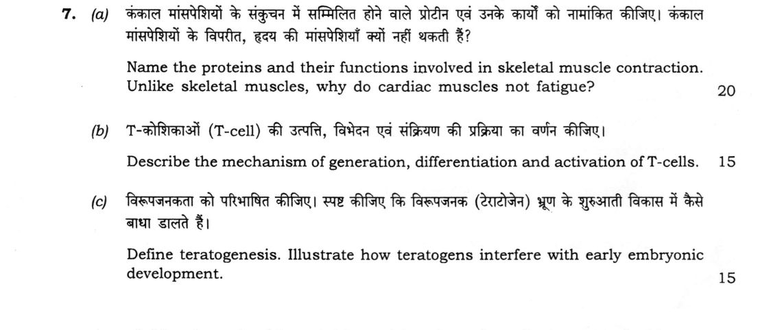
(a) Name the proteins and their functions involved in skeletal muscle contraction. Unlike skeletal muscles, why do cardiac…
Name the proteins and their functions involved in skeletal muscle contraction. Unlike skeletal muscles, why do cardiac muscles not fatigue? 20 marks
Describe the mechanism of generation, differentiation and activation of T-cells. 15 marks
Define teratogenesis. Illustrate how teratogens interfere with early embryonic development. 15 marks
हिंदी में प्रश्न पढ़ें
कंकाल मांसपेशियों के संकुचन में सम्मिलित होने वाले प्रोटीन एवं उनके कार्यों को नामांकित कीजिए। कंकाल मांसपेशियों के विपरीत, हृदय की मांसपेशियाँ क्यों नहीं थकती हैं? (20 अंक)
T-कोशिकाओं (T-cell) की उत्पत्ति, विभेदन एवं संक्रियण की प्रक्रिया का वर्णन कीजिए। (15 अंक)
विरूपजनकता को परिभाषित कीजिए। स्पष्ट कीजिए कि विरूपजनक (टेराटोजेन) भ्रूण के शुरुआती विकास में कैसे बाधा डालते हैं। (15 अंक)
Model answer
Drafted with AI by UPSC Answer Check against this question's marking rubric, to the expected length. UPSC does not publish answers for Mains — this is one way to score well, not an official key. How we build rubrics and answers.
(a) Skeletal muscle proteins, and why cardiac muscle does not fatigue (20 marks)
Proteins of skeletal muscle contraction.
- Contractile: actin, forming the thin filament as a double helix of F-actin, binds the myosin head. Myosin, forming the thick filament, has a tail (light meromyosin) that builds the filament and a head (heavy meromyosin) with ATPase activity that binds actin and pulls it in the power stroke.
- Regulatory: tropomyosin lies along actin and covers the myosin-binding sites at rest. The troponin complex is made of TnT (binds tropomyosin), TnC (binds Ca²⁺) and TnI (inhibits actin–myosin interaction), and Ca²⁺ binding to TnC moves tropomyosin and switches contraction on.
- Structural: titin is an elastic protein anchoring the thick filament to the Z disc and returning the sarcomere to length after stretch. Nebulin sets thin-filament length, α-actinin cross-links actin at the Z disc, myomesin holds the thick filaments at the M line, desmin links adjacent Z discs, and dystrophin links actin to the sarcolemma (its absence causes Duchenne muscular dystrophy).
Why cardiac muscle does not fatigue.
- It is entirely aerobic. Mitochondria take up about a third of the cell volume, and myoglobin is abundant, so ATP is made continuously by oxidative phosphorylation and there is no build-up of lactic acid.
- It has a very rich blood supply, with a capillary for each fibre, and it extracts about 70–75% of the oxygen from blood. It also uses fatty acids, glucose and lactate as fuels.
- Its action potential has a long plateau, so the refractory period (about 250 ms) lasts almost the whole of contraction. It cannot be tetanised, so the heart always relaxes between beats, and the rest during diastole restores perfusion (coronary flow is mainly in diastole).
- The intrinsic pacemaker (SA node) sets a regular rhythm, and intercalated discs and gap junctions make it work as a syncytium.
In skeletal muscle, sustained or intense contraction outstrips the oxygen supply, so it depends on anaerobic glycolysis and fatigues.
(b) Generation, differentiation and activation of T cells (15 marks)
Generation. T-cell precursors arise from haematopoietic stem cells in the bone marrow, pass through common lymphoid progenitors and travel in the blood to the thymus, where they enter as thymocytes.
Differentiation in the thymus.
- Double negative (CD4⁻CD8⁻): the TCR β chain gene is rearranged by V(D)J recombination (RAG1 and RAG2), and pairs with the pre-Tα chain as the pre-TCR. Cells that pass this test (β-selection) proliferate.
- Double positive (CD4⁺CD8⁺): the α chain is rearranged, producing an αβ TCR.
- Positive selection in the cortex: cortical epithelial cells present self peptide–MHC. Cells whose TCR binds self-MHC with moderate affinity survive, and the rest die by neglect. Binding to MHC I gives CD8⁺ cells, and to MHC II gives CD4⁺ cells (single positive).
- Negative selection in the medulla: cells that bind self antigens strongly (with tissue antigens shown by AIRE) undergo apoptosis (central tolerance), and some become regulatory T cells. About 95% of thymocytes die. Mature naive T cells leave for the lymph nodes and spleen.
Activation in the periphery. An antigen-presenting cell (dendritic cell) gives three signals.
- Signal 1: the TCR recognises the peptide–MHC, helped by CD4 or CD8.
- Signal 2: co-stimulation, by CD28 on the T cell binding B7 (CD80/86). Without it, the cell becomes anergic.
- Signal 3: cytokines direct differentiation: IL-12 to Th1, IL-4 to Th2, TGF-β with IL-6 to Th17, TGF-β to Treg, and IL-21 to Tfh.
The activated cells make IL-2 and expand clonally into effector subsets (helper Th1, Th2, Th17, Tfh and Treg, and cytotoxic CD8⁺ cells that kill with perforin and granzymes) and memory cells.
(c) Teratogenesis and how teratogens interfere with early development (15 marks)
Definition. Teratogenesis is the production of congenital malformations by an agent (a teratogen) that disturbs normal embryonic development.
Agents and defects.
- Drugs: thalidomide (limb defects, phocomelia), isotretinoin (retinoic acid; craniofacial and heart defects), valproate (neural tube defects) and warfarin.
- Infections: rubella (cataract, deafness and heart defects), Zika virus (microcephaly), cytomegalovirus and Toxoplasma.
- Chemicals: alcohol (fetal alcohol syndrome), methylmercury, and lead.
- Radiation: ionising radiation (microcephaly).
- Maternal conditions: diabetes (heart defects, caudal regression) and folate deficiency (neural tube defects).
Mechanisms. Teratogens may damage DNA (mutagenesis), kill cells (excess apoptosis), block cell division, migration or differentiation, or disturb signalling and gene expression. Excess retinoic acid upsets Hox gene patterning, thalidomide marks the transcription factor SALL4 for degradation, alcohol kills neural crest cells, and folate antagonists impair DNA synthesis and neural tube closure.
Principles.
- Critical period: sensitivity is greatest during organogenesis (weeks 3–8 in humans), when heart, brain and limbs form. In the first two weeks the effect is all-or-none (death or complete repair), and later exposure causes growth or functional defects.
- Dose: there is a threshold below which no effect is seen, and the severity rises with dose.
- Susceptibility depends on the genotypes of the mother and embryo, and on transfer across the placenta.
How this answer will be evaluated
Evaluation rubric
Each sub-part is marked on its own, against the marks and word limit printed on the paper.
- (a) Name the proteins and their functions involved in skeletal muscle contraction. Unlike skeletal muscles, why do cardiac muscles not fatigue? (20 marks). 20 marks
Must cover
- Contractile proteins actin and myosin with functions
- Regulatory proteins tropomyosin and troponin (TnT, TnC, TnI)
- Structural proteins such as titin, nebulin, α-actinin and dystrophin
- Fatigue-resistance of cardiac muscle: aerobic metabolism, mitochondria and myoglobin, rich blood supply, long refractory period
Loses marks
- Only naming actin and myosin.
- Stating that cardiac muscle is anaerobic.
Earns more
- Function stated for each protein.
- Cardiac reasoning given, not a list of features.
Extra mark
- Intercalated discs and SA node rhythm
- (b) Describe the mechanism of generation, differentiation and activation of T-cells (15 marks). 15 marks
Must cover
- Origin in bone marrow and entry into the thymus
- Thymic differentiation: TCR gene rearrangement, DN to DP to SP
- Positive and negative selection with central tolerance
- Peripheral activation by three signals and effector subsets
Loses marks
- Confusing T cell with B cell development.
- Omitting selection.
Earns more
- Named molecules such as RAG, CD28–B7 and AIRE.
- Th subsets with their cytokines.
Extra mark
- Memory cells and Treg
- (c) Define teratogenesis. Illustrate how teratogens interfere with early embryonic development (15 marks). 15 marks
Must cover
- Definition of teratogenesis
- Named teratogens and specific defects
- Mechanisms: DNA damage, cell death, disturbed signalling
- Critical period and dose dependence
Loses marks
- Defining without illustration.
- Confusing teratogens with mutagens only.
Earns more
- Example of thalidomide or rubella with the defect.
- Weeks of organogenesis stated.
Extra mark
- All-or-none period and genotype of mother and embryo
What "Explain" is asking you to do
Make the working of something clear — what sets it off, what follows from what, and what it produces. Explain is the Commission's mechanism word: it dominates the technical papers and the “explain why” stems, where the marks sit in the causal chain and not in the label.
Structure that answers it
State what it is → the initiating condition → the chain of cause, step by step → an instance where it plays out → what the chain produces
Where marks are lost
Describing what something looks like instead of why it works that way. Naming the stages without linking them reads as description too.
Practice this exact question
Write your answer and it is marked point by point against the model answer above — what you covered, what you missed, what you got wrong.
Evaluate my answer →More from Zoology 2026 Paper II
- Q4 (a) What is Oparin concept? Comment on the chemical theory of origin of life. (20 marks)…
- Q5 Write on the following in about 150 words each : (a) Role of calcium in muscle contractio…
- Q6 (a) Explain various levels of protein organization with suitable examples. (20 marks) (b)…
- Q7 (a) Name the proteins and their functions involved in skeletal muscle contraction. Unlike…
- Q8 (a) Define apoptosis. Explain the characteristics, molecular pathways and significance of…