Paper II — Q6
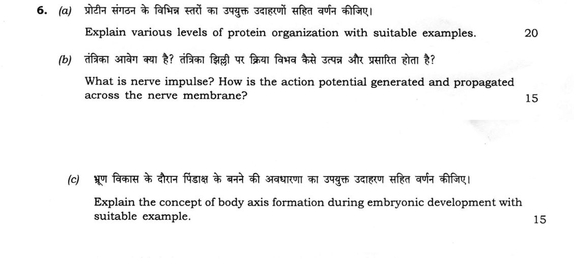
(a) Explain various levels of protein organization with suitable examples. (20 marks) (b) What is nerve impulse? How is the…
Explain various levels of protein organization with suitable examples. 20 marks
What is nerve impulse? How is the action potential generated and propagated across the nerve membrane? 15 marks
Explain the concept of body axis formation during embryonic development with suitable example. 15 marks
हिंदी में प्रश्न पढ़ें
प्रोटीन संगठन के विभिन्न स्तरों का उपयुक्त उदाहरणों सहित वर्णन कीजिए। (20 अंक)
तंत्रिका आवेग क्या है? तंत्रिका झिल्ली पर क्रिया विभव कैसे उत्पन्न और प्रसारित होता है? (15 अंक)
भ्रूण विकास के दौरान पिंडाक्ष के बनने की अवधारणा का उपयुक्त उदाहरण सहित वर्णन कीजिए। (15 अंक)
Model answer
Drafted with AI by UPSC Answer Check against this question's marking rubric, to the expected length. UPSC does not publish answers for Mains — this is one way to score well, not an official key. How we build rubrics and answers.
(a) Levels of protein organisation with examples (20 marks)
Proteins have four levels of organisation.
- Primary structure: the linear sequence of amino acids in the polypeptide chain, joined by covalent peptide bonds from the N-terminus to the C-terminus. The sequence is set by the gene and determines all higher levels. Example: insulin, with an A chain of 21 residues and a B chain of 30, joined by disulphide bonds, whose sequence was worked out by Sanger.
- Secondary structure: regular local folding of the backbone, held by hydrogen bonds between the C=O and N–H groups of the peptide bonds. The α-helix is a right-handed coil with 3.6 residues per turn, as in keratin and in myoglobin. The β-pleated sheet, parallel or antiparallel, is found in silk fibroin. Others are β-turns and the collagen triple helix (glycine at every third position).
- Tertiary structure: the overall three-dimensional folding of one polypeptide, held by interactions among the side chains: hydrophobic interactions (the main force, burying non-polar groups in the core), hydrogen bonds, ionic bonds (salt bridges), van der Waals forces and covalent disulphide bonds. Example: myoglobin, a compact globular chain of 153 residues, mostly α-helix, with one haem group, and lysozyme and ribonuclease. Supersecondary motifs and domains are units within it.
- Quaternary structure: the association of two or more polypeptide subunits, held by the same non-covalent forces (and sometimes disulphide bonds). Example: haemoglobin, a tetramer (α₂β₂) that binds oxygen cooperatively; other examples are lactate dehydrogenase and immunoglobulins.
Note. Heat, extreme pH or urea denature a protein by disrupting the higher levels, without breaking peptide bonds, and it loses its function. Anfinsen showed with ribonuclease that the primary structure carries the information for folding, and chaperones assist.
(b) Nerve impulse, and generation and propagation of the action potential (15 marks)
Nerve impulse. A nerve impulse is a wave of electrochemical change, a self-propagating depolarisation, that travels along the neuron membrane.
Resting potential. The inside of the axon is negative, at about −70 mV, because of the Na⁺–K⁺ pump (3 Na⁺ out and 2 K⁺ in) and the greater leak of K⁺ than Na⁺.
Generation of the action potential.
- A stimulus depolarises the membrane, and when the threshold (about −55 mV) is reached, voltage-gated Na⁺ channels open. Na⁺ rushes in, and depolarisation is rapid, with an overshoot to about +30 mV.
- The Na⁺ channels then inactivate, and the voltage-gated K⁺ channels open more slowly. K⁺ moves out and the membrane repolarises, with a brief hyperpolarisation, and the pump restores the ion distribution.
- It is all-or-none: a stimulus below threshold gives no impulse, and a larger one does not raise the size of the spike.
- In the absolute refractory period (Na⁺ channels inactivated) no impulse can be started, and in the relative refractory period only a strong stimulus can. This makes the impulse move in one direction.
Propagation. The depolarised region sets up local circuit currents to the neighbouring resting region, which is brought to threshold, and so the impulse travels along.
- In unmyelinated fibres the conduction is continuous, at about 0.5–2 m/s.
- In myelinated fibres it is saltatory: the impulse jumps between nodes of Ranvier, where Na⁺ channels are concentrated, at up to about 100 m/s, faster and using less energy.
- The velocity increases with axon diameter, myelination and temperature. Demyelination in multiple sclerosis slows conduction.
(c) Body axis formation in embryonic development (15 marks)
Concept. During early development the embryo establishes its anteroposterior (head–tail), dorsoventral (back–belly) and left–right axes. The axes are set by localised maternal determinants (mRNAs and proteins in the egg), by gradients of morphogens, and by organiser tissue that induces neighbouring cells. Axis information then switches on regional genes (the Hox genes).
Example: Drosophila.
- Anteroposterior axis: bicoid mRNA, placed at the anterior pole by nurse cells, is translated into a Bicoid protein gradient (highest anteriorly) that switches on hunchback and head genes. Nanos mRNA at the posterior end forms a Nanos gradient that blocks hunchback translation, so the posterior develops. Gap, pair-rule, segment-polarity and Hox (homeotic) genes then divide the embryo into segments.
- Dorsoventral axis: the Toll receptor is activated ventrally by Spätzle, so that a gradient of nuclear Dorsal protein forms (highest ventrally) and switches on twist and snail (mesoderm), with dpp dorsally.
Example: Xenopus.
- After fertilisation, cortical rotation moves the cortex about 30°, so the dorsal side forms opposite the sperm entry point. β-catenin (Wnt pathway) accumulates on the dorsal side and sets up the Nieuwkoop centre in dorsal vegetal cells.
- This induces the Spemann–Mangold organiser in the dorsal lip of the blastopore. The organiser secretes noggin, chordin and follistatin, which block BMP-4 and dorsalise the mesoderm and induce the neural plate. Spemann and Mangold (1924) showed that a grafted dorsal lip produced a second axis.
- The left–right axis arises from the leftward flow of fluid driven by cilia in the node, which switches on Nodal and Pitx2 on the left.
How this answer will be evaluated
Evaluation rubric
Each sub-part is marked on its own, against the marks and word limit printed on the paper.
- (a) Explain various levels of protein organisation with suitable examples (20 marks). 20 marks
Must cover
- Primary structure: amino acid sequence and peptide bonds, with an example such as insulin
- Secondary structure: α-helix and β-sheet with hydrogen bonds, and examples
- Tertiary structure: side-chain interactions and disulphide bonds, with myoglobin
- Quaternary structure: subunits, with haemoglobin
Loses marks
- Naming levels without examples.
- Assigning peptide bonds to secondary structure.
Earns more
- Bonds that stabilise each level named.
- Denaturation and Anfinsen's experiment.
Extra mark
- Domains, motifs and chaperones
- (b) What is nerve impulse? How is the action potential generated and propagated across the nerve membrane? (15 marks). 15 marks
Must cover
- Nerve impulse defined, with the resting potential and the Na⁺–K⁺ pump
- Generation: threshold, voltage-gated Na⁺ opening, depolarisation, K⁺ repolarisation, all-or-none
- Refractory periods and their role
- Propagation by local circuits, continuous and saltatory conduction, and factors affecting velocity
Loses marks
- Confusing action potential with resting potential.
- Omitting propagation.
Earns more
- Numerical values of resting and peak potentials.
- Role of nodes of Ranvier.
Extra mark
- Clinical example such as multiple sclerosis
- (c) Explain the concept of body axis formation during embryonic development with suitable example (15 marks). 15 marks
Must cover
- Concept: anteroposterior, dorsoventral and left–right axes set by maternal determinants, morphogen gradients and organisers
- Drosophila: bicoid and nanos gradients, and Dorsal gradient with Toll
- Xenopus: cortical rotation, β-catenin, Nieuwkoop centre and the Spemann–Mangold organiser
- Left–right axis and Hox genes
Loses marks
- Describing only cleavage.
- Naming axes with no mechanism.
Earns more
- At least one model system in detail.
- Molecules such as noggin, chordin and BMP-4.
Extra mark
- Spemann and Mangold's graft experiment
What "Explain" is asking you to do
Make the working of something clear — what sets it off, what follows from what, and what it produces. Explain is the Commission's mechanism word: it dominates the technical papers and the “explain why” stems, where the marks sit in the causal chain and not in the label.
Structure that answers it
State what it is → the initiating condition → the chain of cause, step by step → an instance where it plays out → what the chain produces
Where marks are lost
Describing what something looks like instead of why it works that way. Naming the stages without linking them reads as description too.
Practice this exact question
Write your answer and it is marked point by point against the model answer above — what you covered, what you missed, what you got wrong.
Evaluate my answer →More from Zoology 2026 Paper II
- Q3 (a) What is recombinant DNA technology? Describe the different vectors used in DNA clonin…
- Q4 (a) What is Oparin concept? Comment on the chemical theory of origin of life. (20 marks)…
- Q5 Write on the following in about 150 words each : (a) Role of calcium in muscle contractio…
- Q6 (a) Explain various levels of protein organization with suitable examples. (20 marks) (b)…
- Q7 (a) Name the proteins and their functions involved in skeletal muscle contraction. Unlike…
- Q8 (a) Define apoptosis. Explain the characteristics, molecular pathways and significance of…